
Image
|
Figure Caption
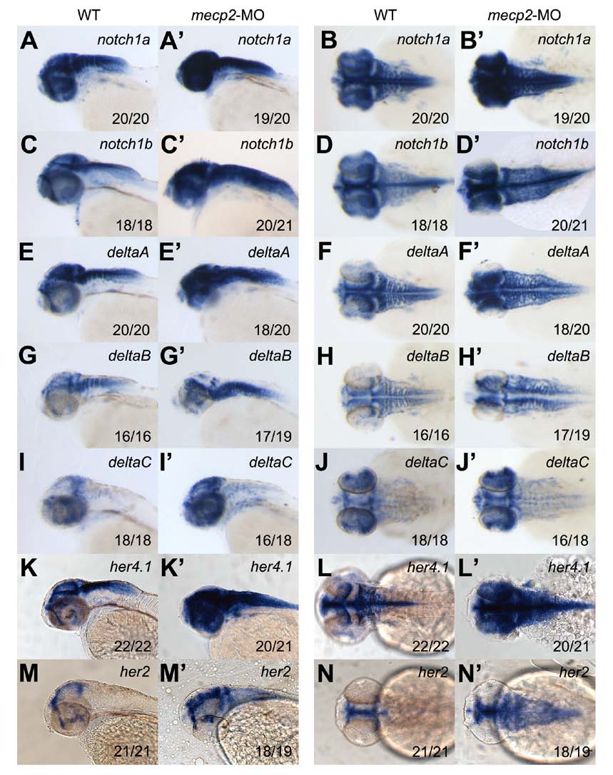
Fig. S1 Knockdown of mecp2 enhances Notch signaling activity in the developing brain. RNA in situ hybridization with notch1a, notch1b, deltaA, deltaB, deltaC, her4.1, and her2 probes in controls (A-J) and mecp2 morphants (A′-J′) at 48 hpf. Note up-regulated expression of these Notch signaling genes in the developing brain of mecp2 morphants. Lateral views (A-A′; C-C′; E-E′; G-G′; I-I′; K-K′; M-M′) and dorsal views (B-B′; D-D′; F-F′; H-H′; J-J′; L-L′; N-N′) with the anterior to the left. The numbers in the bottom right corner represent phenotypic embryos/total embryos.
Acknowledgments
This image is the copyrighted work of the attributed author or publisher, and
ZFIN has permission only to display this image to its users.
Additional permissions should be obtained from the applicable author or publisher of the image.
Full text @ J. Cell Sci.