
|
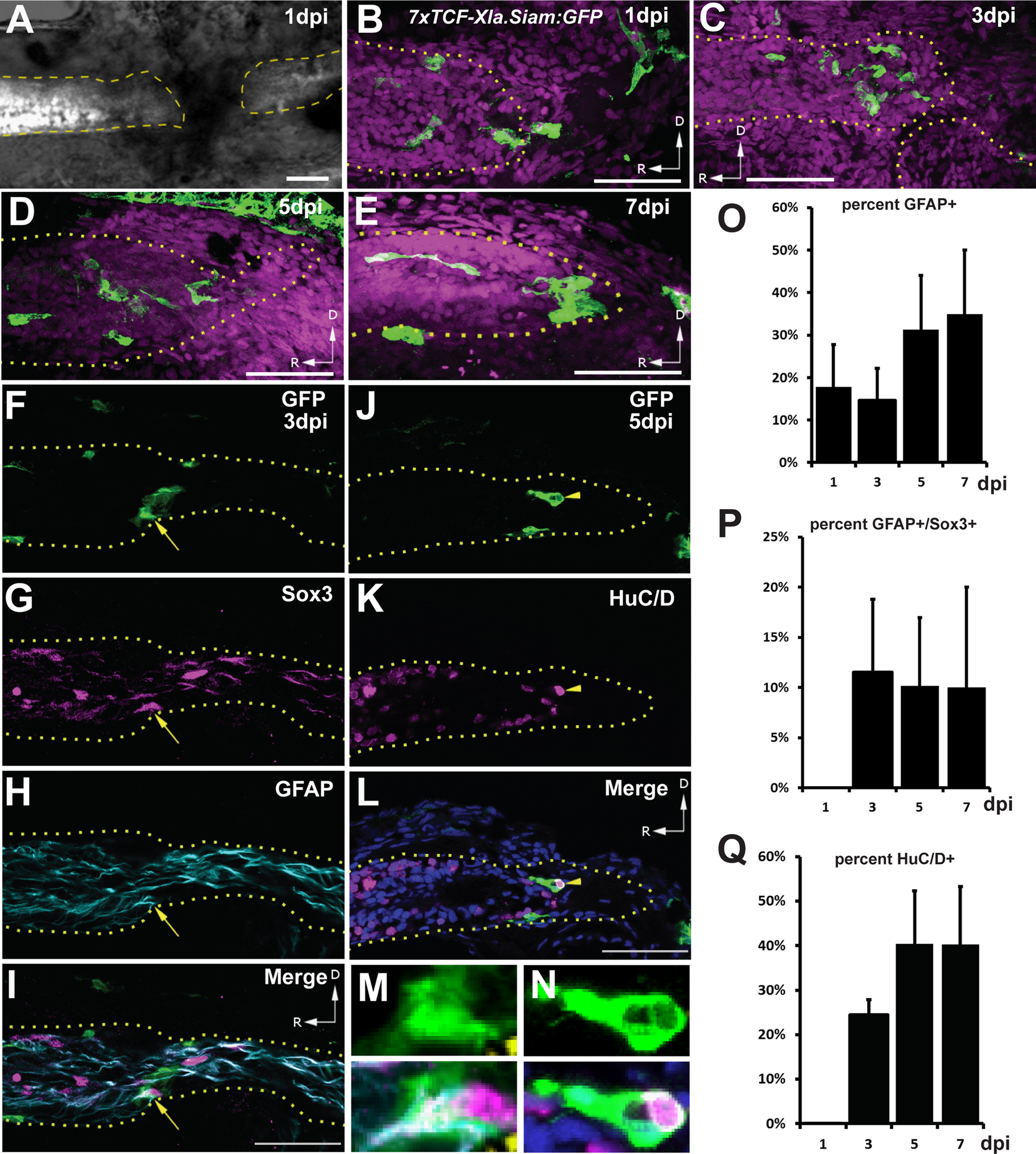
Fig. 1
Wnt reporter expression after SCI. (A) Lateral view of a transected spinal cord at 1 dpi showing loss of neurons (white, labeled by elavl3:EGFP) at the injury site. (B–E) The 7xTCF-Xla.Siam:GFP Wnt reporter (green) is expressed in the regeneration blastema from 1 to 7 dpi. Magenta channel is nuclear staining. (F–I) Wnt reporter-expressing cells express GFAP and Sox3 at 3 dpi (arrow). (J–L) Other reporter-expressing cells are HuC/D+ neurons at 5 dpi (arrowhead). Blue channel is nuclear staining. (M,N) High-magnification views of co-labeled cells from panels (I and L). Percentage of GFP+ cells co-expressing GFAP, GFAP and Sox3, and HuC/D. Image in (A) is merged from fluorescent and brightfield channels obtained with a compound microscope. Images in (B–N) are single confocal slices from lateral views of whole-mounted larvae. Scalebars=50 µm, dashed/dotted lines outline the spinal cord, and D/R arrows indicate dorsal and rostral, respectively. For (O–Q), n=5 sections at each timepoint; error bars=SEM.
Reprinted from Developmental Biology, 403(1), Briona, L.K., Poulain, F.E., Mosimann, C., Dorsky, R.I., Wnt/ß-catenin signaling Is required for radial glial neurogenesis following spinal cord injury, 15-21, Copyright (2015) with permission from Elsevier. Full text @ Dev. Biol.