
|
Fig. 6
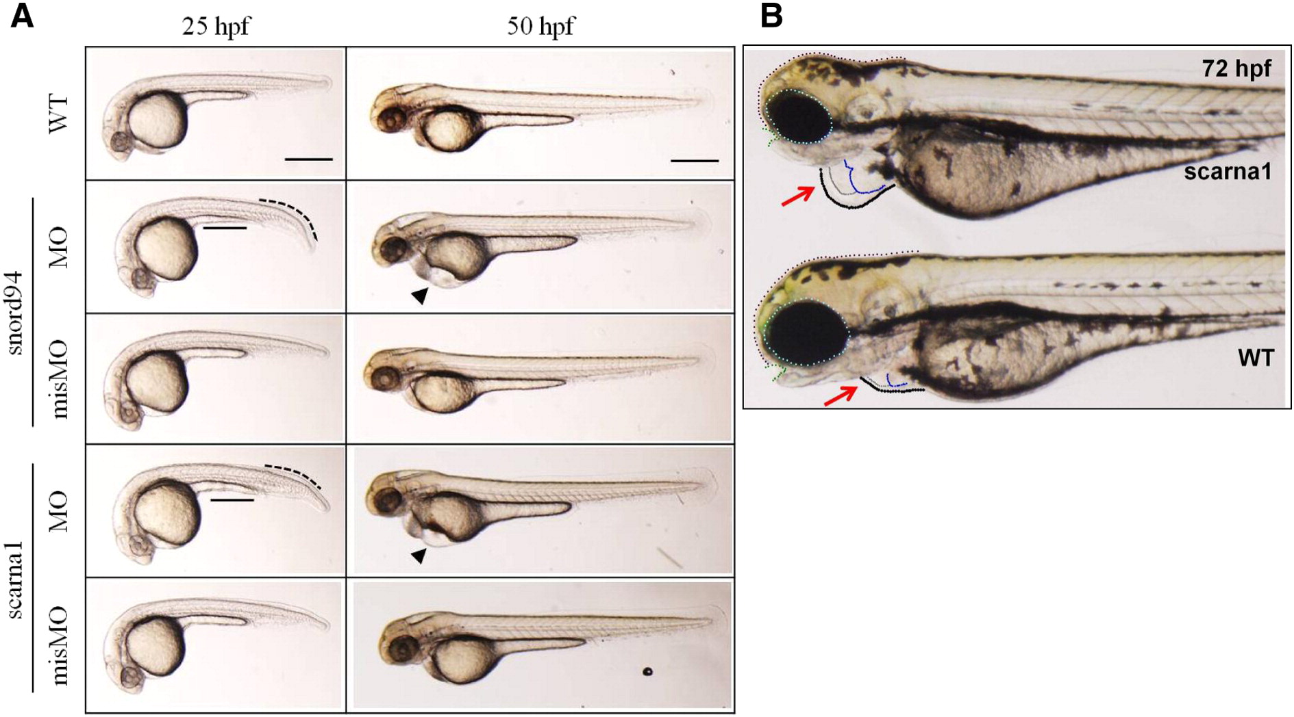
Heart deformities in MO-injected zebrafish. (A) Lateral views of wild-type and MO-injected embryos at 25 and 50 hpf. Both the snord94 and scarna1 morphants display little developmental delay with bent tail (black dotted curved line) and improperly formed yolk extension (black solid line) at 25 hpf. The pericardial edema (black triangle) and deformed yolk sac were more evident at 50 hpf. (B) Enlarged images of the heart region in wild-type and MO-injected embryos at 72 hpf. The conspicuous pericardial edema (black dotted circle) was observed in snoRNA MO-injected embryos. Gray outline is the atrium, blue outline is the ventricle. Scale bars: 200 µm.
Reprinted from Biochimica et biophysica acta. Molecular basis of disease, 1852(8), Patil, P., Kibiryeva, N., Uechi, T., Marshall, J., O'Brien, J.E., Artman, M., Kenmochi, N., Bittel, D.C., scaRNAs Regulate Splicing and Vertebrate Heart Development, 1619-29, Copyright (2015) with permission from Elsevier. Full text @ BBA Molecular Basis of Disease