
|
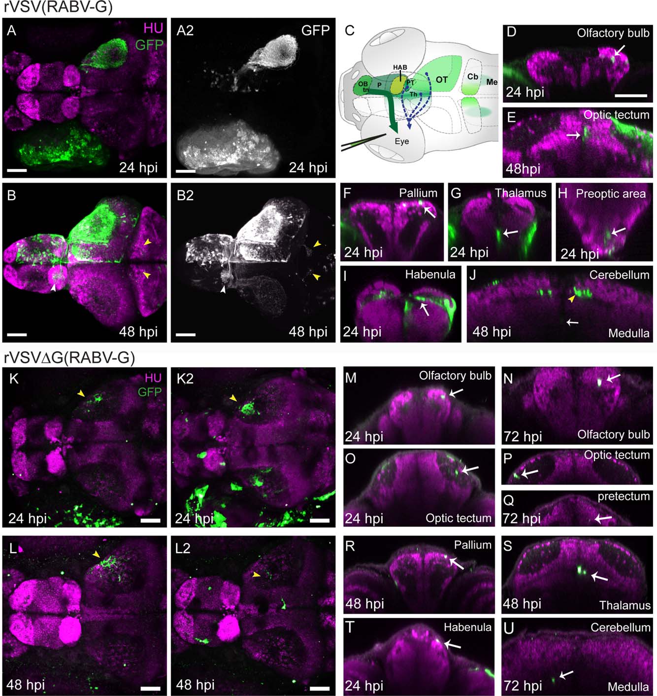
Fig. 7
Labeling of centrifugal circuits in zebrafish with rVSV(RABV-G). (A,B) Dorsal view of confocal maximal projections showing rVSV(RABV-G) labeling (green, GFP) and HU (magenta, pan-neuronal marker) at 24 hpi (A) and 48 hpi (B). GFP channel from (A,B), with signal enhanced to show cells in the brain, is shown in (A2,B2). Images in (B-B2) were captured after dissection of the brain from surrounding tissues. GFP-labeled axonal projections from the contralateral to the ipsilateral habenula (white arrowhead) and projections from the OT to the cerebellum (yellow arrowheads) are indicated in (B-B2). (C) Illustration showing injection of rVSV(RABV-G) into the eye of 3 dpf larval zebrafish. Centrifugal axons in the terminal nerve (tn) originate from the contralateral olfactory bulb (OB) and ventral pallium (P), and project to target cells in the retina (dark green arrow). The locations of rVSV(RABV-G)-infected cells after unilateral eye injection is indicated by green shaded areas. Projections from other centrifugal neurons previously reported among teleost fish are indicated by dashed arrows from the thalamus (Th), OT, and pretectum (PT). (D-J) Transverse optical sections from 24 hpi or 48 hpi are shown. At 24 hpi, labeling is restricted to the RGC axons in the neuropil of OT, and cell bodies in the OB (D), OT (E, 48 hpi shown), pallium (F), thalamus (G), preoptic area (H), habenula (I), medulla (J, 48 hpi shown), and pretectum (not shown). At 48 hpi, additional labeling was seen, including GFP-positive efferent axons (yellow arrowhead) projecting into the contralateral and ipsilateral cerebellum (J). (K-L2) Confocal maximal projections of GFP expression and HU after injection of 1 × 10⁸ ffu/mL (K,L), or 1 × 10⁹ ffu/mL (K2,L2) rVSVΔG(RABV-G), at 24-48 hpi. Yellow arrowheads in (K-L2) indicate RGC axons in the neuropil of the OT. In zebrafish injected with the higher dose of rVSVΔG(RABV-G), labeling revealed primary infection in centrifugal neurons. (M-U) Representative transverse optical sections from 24-72 hpi show the results of centrifugal labeling, as sparse GFP-expressing cells in the OB (M,N), OT (O,P), pretectum (Q), pallium (R), thalamus (S), habenula (T), and medulla (U). GFP-labeled cells were not detected in the preoptic area (not shown) and labeled axons were not present in the cerebellum (U) following infections with rVSVΔG (RABV-G). CB = cerebellum, HAB = habenula, Me = medulla, OB = olfactory bulb, OT = optic tectum, P= pallium, PT = pretectum, PO = preoptic area, tn = terminal nerve, Scale bars: = 50 µm.