
Fig. 3
- ID
- ZDB-IMAGE-150716-16
- Genes
- Publication
- Xiao et al., 2015 - High-resolution live imaging reveals axon-glia interactions during peripheral nerve injury and repair in zebrafish
- All Figures
- Figures for Xiao et al., 2015
|
Fig. 3
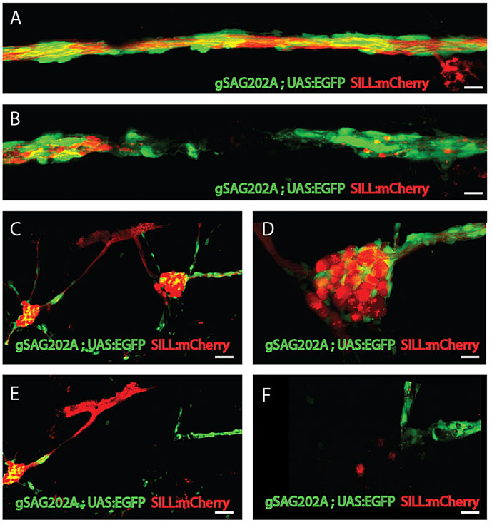
Selective axonal severing and neuronal ablation by laser>. (A,B) High magnification of the lateral line nerve (A) before laser ablation and (B) after laser ablation. The nerve and Schwann cells are precisely and completely severed with no visible damage to the surrounding tissue. (C) The maximal projections of anterior and posterior ganglion before ablation. (D) Higher magnification of posterior ganglion before ablation. (E) The image of the same animal after ablation reveals that the posterior ganglion was totally ablated. (F) Higher magnification of posterior ganglion after ablation. Scale bars: 10µm (A,B,D,F) and 50µm (C,E).