
|
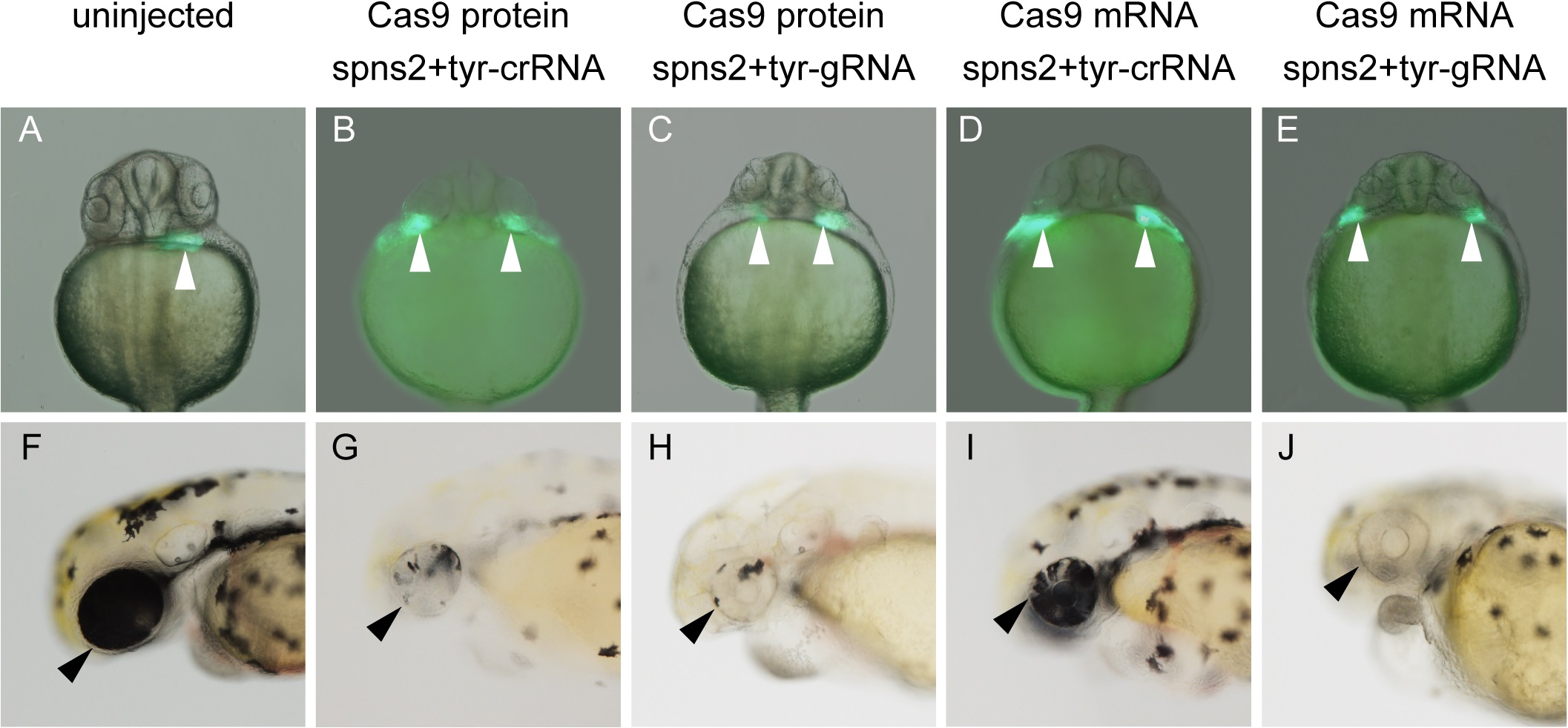
Fig. 1
Phenotypic analysis in the embryos injected with two crRNAs, tracrRNA and Cas9 protein.
(A, F) An uninjected embryo derived from Tg(cmlc2:eGFP) expressing eGFP in the cardiac cells. (B, G) Tg(cmlc2:eGFP)-derived embryos injected with spns2-crRNA1 (25 pg), tyr-crRNA (25 pg), tracrRNA (100 pg) and Cas9 protein (400 pg). (C, H) Tg(cmlc2:eGFP)-derived embryos injected with spns2-gRNA1 (25 pg), tyr-gRNA (25 pg) and Cas9 protein (400 pg). (D, I) Tg(cmlc2:eGFP)-derived embryos injected with spns2-crRNA1 (25 pg), tyr-crRNA (25 pg), tracrRNA (100 pg) and Cas9 mRNA (250 pg). (E, J) Tg(cmlc2:eGFP)-derived embryos injected with spns2-gRNA1 (25 pg), tyr-gRNA (25 pg) and Cas9 mRNA (250 pg). (A-E) The injection of the two crRNAs and tracrRNA with Cas9 protein or Cas9 mRNA (B and C) as well as the injection of two gRNAs with Cas9 protein or Cas9 mRNA (D and E) caused the cardiac progenitor migration defects at 1 day post-fertilization (dpf), whereas an uninjected embryo had a normal heart (A). White arrowheads indicate the position of the developing heart. (F–J) The injection of the two crRNAs and tracrRNA with Cas9 protein or Cas9 mRNA (G and H) as well as the injection of two gRNAs with Cas9 protein or Cas9 mRNA (I and J) caused pigmentation defects in the retinal epithelium at 2 dpf, whereas an uninjected embryo had retinal epithelial cells with normal pigmentation (F). Black arrowheads indicate the position of the eye. The embryos in (A), (B), (C), (D) and (E) correspond to the embryos in (F), (G), (H), (I) and (J), respectively. (A-E) Ventral view with anterior at the top. (F-J) Lateral view with anterior to the left and dorsal at the top.