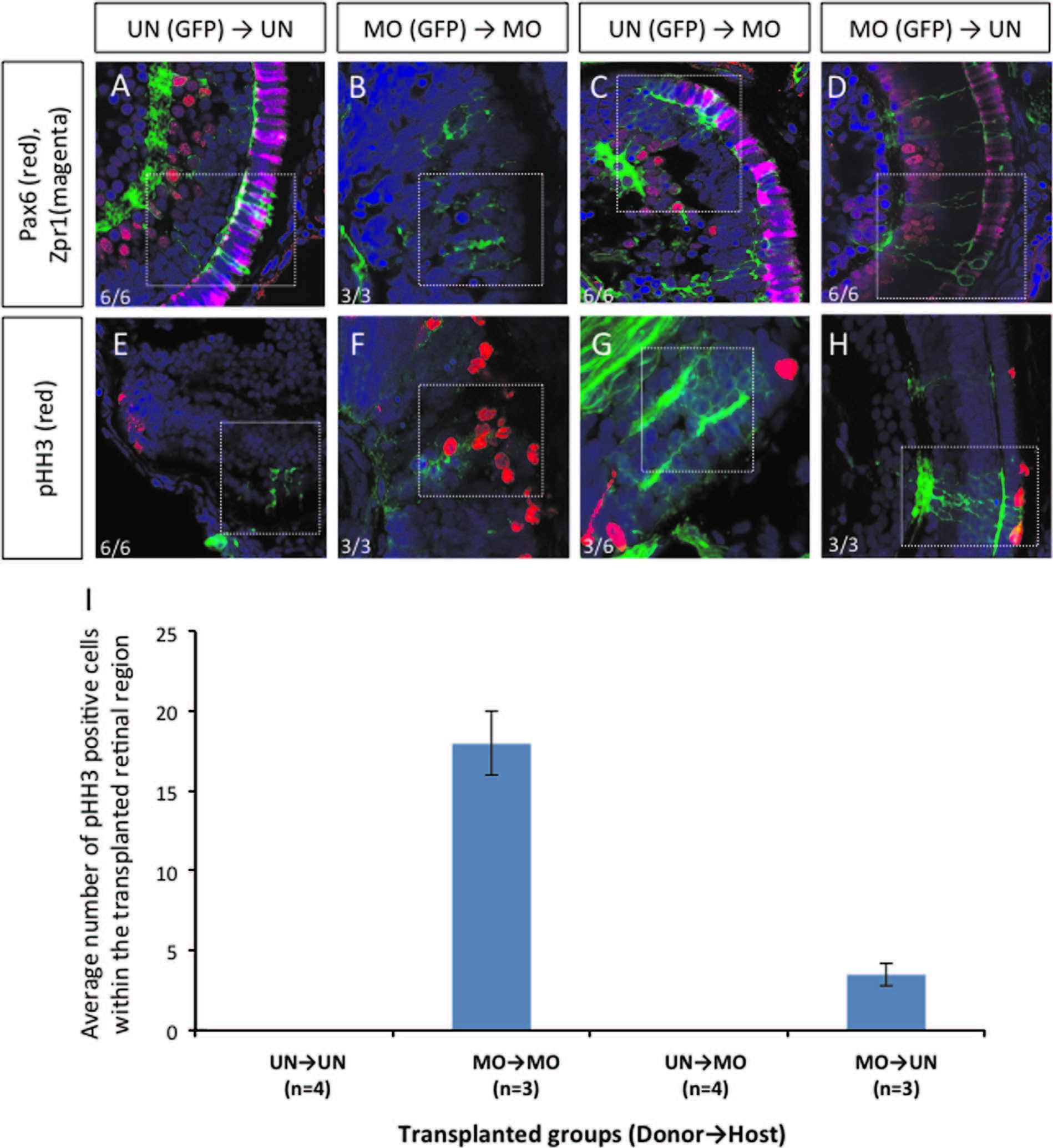
Fig. 1
Transplantation experiments demonstrated that dmbx1 functions cell autonomously in the developing retina. Coronal sections of 72 hpf host embryos transplanted with β-actin:GFP positive donor cells at blastula stage. Cells from un-injected (UN) or morpholino-injected (MO) Tg(β-actin:GFP) donor embryos were transplanted to either UN- or MO host embryos. Animals with mosaic retinae were selected for further analysis using immunohistochemistry: Pax6 (RGCs and amacrine cells) and Zpr1 (cone photoreceptor) (A–D, n=6 in each UN donor group and n=3 in each MO donor group); and phospho-Histone H3 [pHH3] (E–H, n=6 in each UN donor group and n=3 in each MO donor group). Images were captured using a confocal microscope without any maximum projection. White dotted boxes highlight areas where GFP+ donor cell localization in the host retina. Numbers of pHH3+ cells within those transplanted cells in the retina of the host embryos are summarized in graph (I).
Reprinted from Developmental Biology, 402(2), Wong, L., Power, N., Miles, A., Tropepe, V., Mutual antagonism of the paired-type homeobox genes, vsx2 and dmbx1, regulates retinal progenitor cell cycle exit upstream of ccnd1 expression, 216-28, Copyright (2015) with permission from Elsevier. Full text @ Dev. Biol.

