
Image
|
Figure Caption
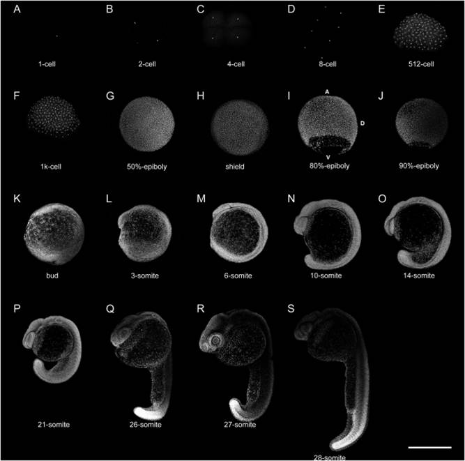
Fig. 2
Nuclei distribution patterns during early zebrafish development. DNA and nuclei were visualized in whole SYTOX Green stained embryos by laser confocal microscopy. The images in this figure are complementary to images shown in Fig. 1. Scale bar 500 µm.
Acknowledgments
This image is the copyrighted work of the attributed author or publisher, and
ZFIN has permission only to display this image to its users.
Additional permissions should be obtained from the applicable author or publisher of the image.
Full text @ Anat. Rec. (Hoboken)