
|
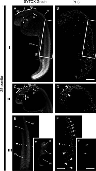
Fig. 13
Proliferation patterns in 28-somite stage embryos. I: Full confocal slice projections of an entire embryo. II: Partial confocal slice projections of the brain region. White doted lines highlight structure like the eye (ey), midbrain (mb), cerebellum (c), hindbrain (hb), and otic vesicle (ov). White arrowheads in D indicate mitotic nuclei in midbrain and hindbrain. White arrows indicate regions with less abundant mitotic nuclei in brain regions. III: Partial projections of the tail region show structures corresponding to the notochord (nc) and myotomes (my). (e and f) Inserts show sagital sections of images indicated in images E, F by the white dotted lines. Notice the positive nuclei for PH3, except in the corresponding notochord and yolk extension. ey, eye; mb, midbrain; hb, hindbrain; c, cerebellum; nt, neural tube; ye, yolk extension. 200 µm scale bar in B. 100 µm scale bar in f insert.