
|
Fig. 3
Molecular Tattooing Unveils the Autonomous Function of Non-muscle Myosin 2 in the Migration of pLLp of Live Zebrafish Embryos
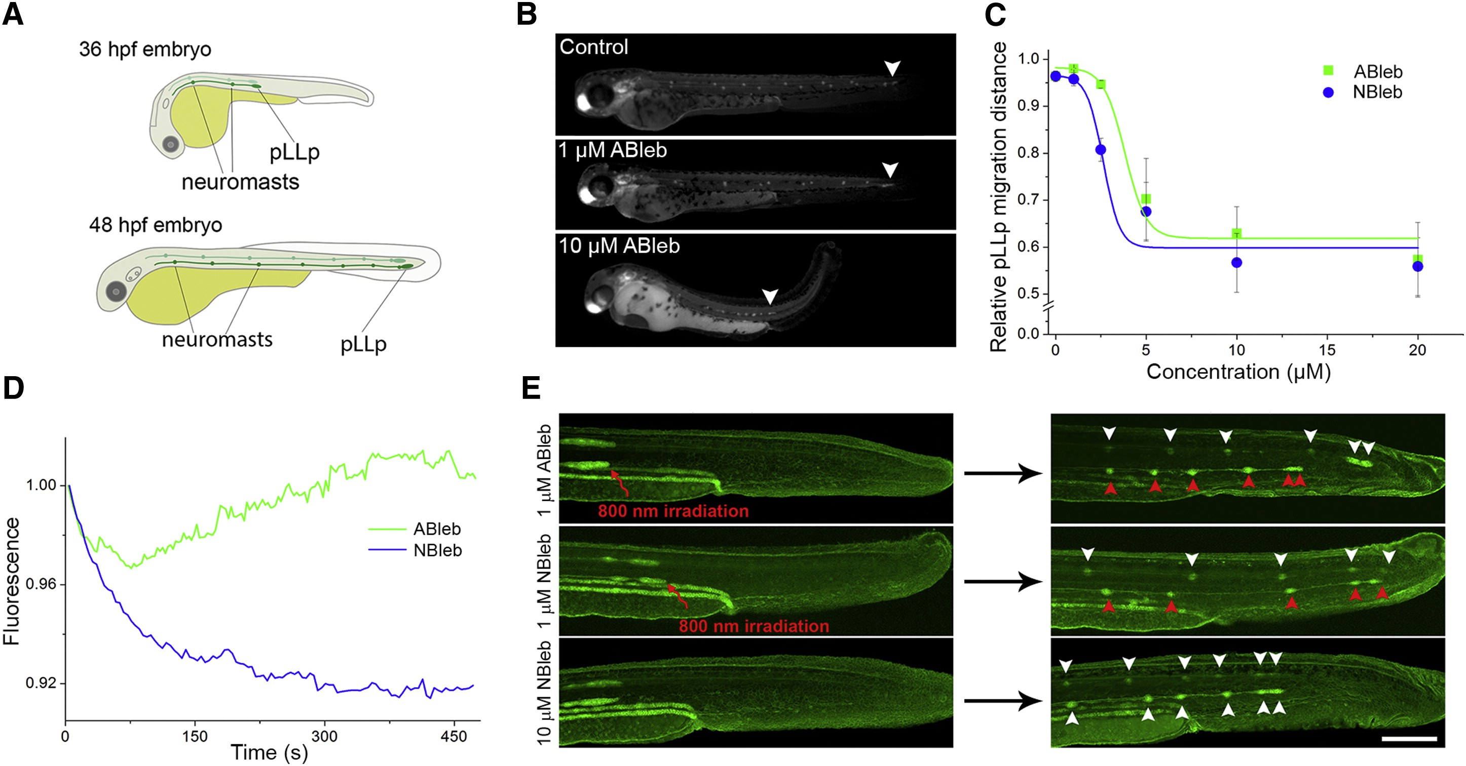
(A) Schematic drawings of zebrafish embryos displaying the spatial formation of the lateral line.
(B) Fluorescence images of 48 hpf zebrafish embryos incubated in the absence and presence of ABleb in dark for 24 hr. White arrowheads mark the position of the halted pLLps.
(C) Relative distance of pLLp migration in the presence of ABleb or NBleb (1 = distance from the otic vesicle to the tip of the tail). Dose-response curves were fitted to means ± SD (n = 4).
(D) Relative fluorescence change of pLLps in live cldn:gfp zebrafish embryo during 2P irradiation in the presence of ABleb or NBleb.
(E) Projected confocal z-stack images of zebrafish embryos with 2P-irradiated pLLps on one side (indicated by red arrows) in the presence of 1 µM ABleb, 1 µM NBleb, and non-irradiated embryo in the presence of 10 µM NBleb. White (non-irradiated) and red (irradiated) arrowheads mark sites of neuromast deposition. See also Movie S1. Scale bar represents 200 µm.