
|
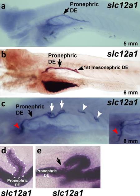
Fig. 8
Expression of slc12a1 in the DE. A–C: Whole mount in situ hybridizations of larvae show that the first mesonephric DE forms on top of the pronephric DE and expresses slc12a1 at the 6 mm stage (B arrowhead). At the 8 mm stage, several more DEs appear on top of the pronephric DE (C white arrows) and pronephric DL (C white arrowheads), and in the rostral region of the pronephric DE (C red arrowhead and inset). The inset is a magnified image of the red arrowhead at a different angle. D: A magnified image of a branch point between the mesonephric DE (dashed line) and the pronephric DE. E: At the 8 mm stage, slc12a1 is downregulated at the junction point with the pronephric DE. DE: distal early segment; DL: distal late segment.