
Fig. 4
- ID
- ZDB-IMAGE-150601-8
- Publication
- Benini et al., 2015 - slc7a6os Gene Plays a Critical Role in Defined Areas of the Developing CNS in Zebrafish
- All Figures
- Figures for Benini et al., 2015
|
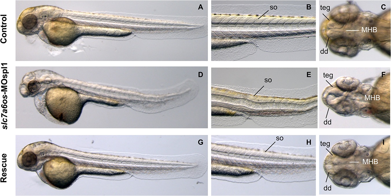
Fig. 4 Morphological analysis of slc7a6os morphants and phenotypic rescue at 48 h.
At 48 hpf, lateral view show that MO injected embryos have an evident pericardial and yolk-sac edema and the somite structures are not well-defined (D-E) when compared to controls (A, D). Dorsal view of morphants shows that the CNS abnormalities are more pronounced and reveals a severe disorganization of the following structures: midbrain–hindbrain boundary, dorsal diencephalon and tegmentum (F). Also at this developmental stage the expression of synthetic slc7a6os mRNA rescues the wild type phenotype in morphants (G-I). Abbreviations: so, somites; teg, tegmentum; dd, dorsal diencephalon; MHB, midbrain–hindbrain boundary.