
|
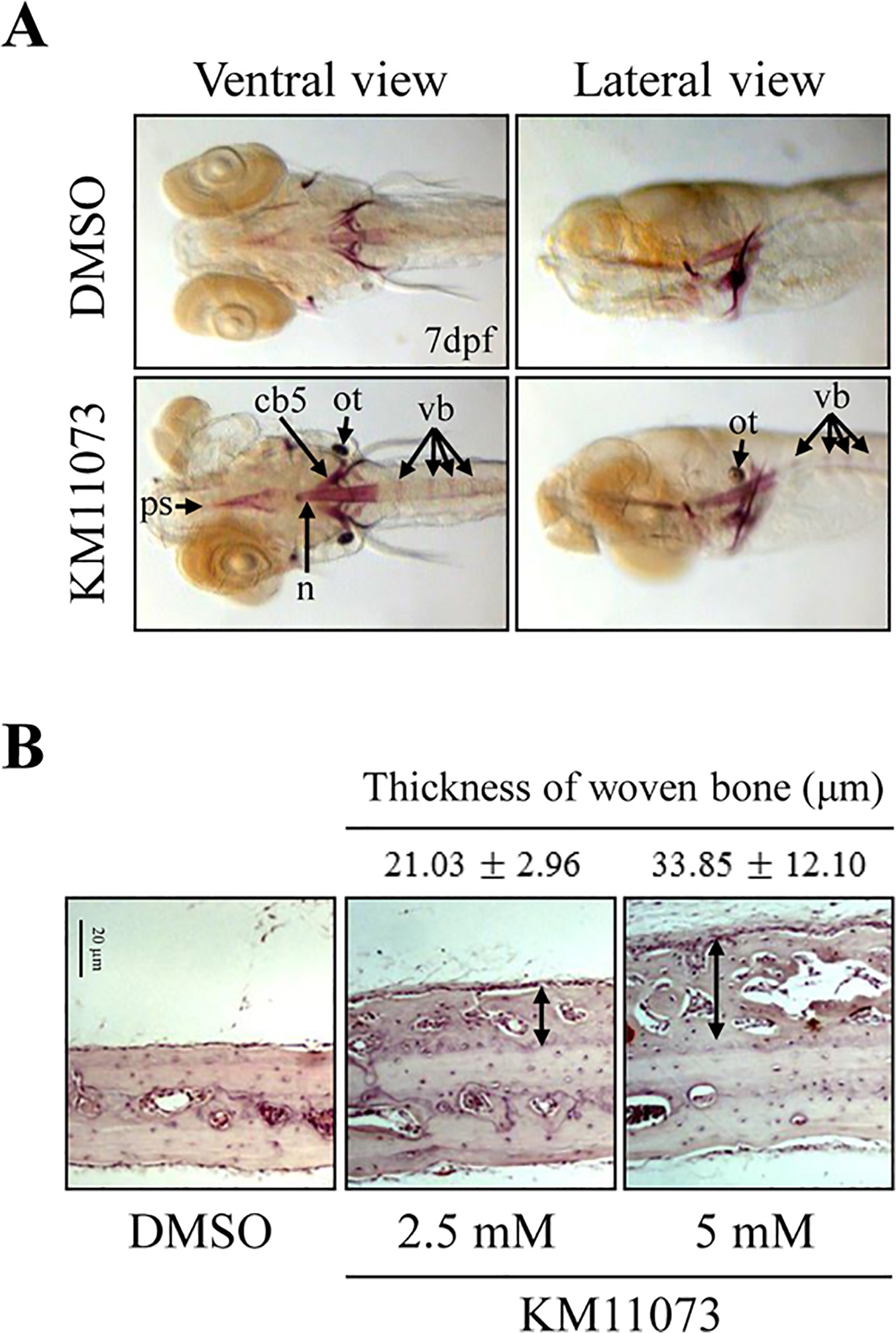
Fig. 5 Evaluation of the in vivo osteogenic activity of KM11073 in zebrafish and mouse calvariae.
Five days after fertilization, zebrafish were treated with KM11073 (1 µM) for 2 days and then fixed and stained with alizarin red S. The parasphenoid (ps), notochord (n), ceratobranchial 5 (cb5), otolith (ot), and vertebrae (vb) are indicated with arrows (A). Collagen sponges soaked in 5 µl of 2.5 or 5 mM KM11073 were placed onto mouse calvarial bones. After 3-week implantation, the mice were sacrificed. Calvarial bones were removed, fixed, decalcified, embedded in paraffin, and sectioned. Sections were stained with H&E and photographed at 200 × magnification. Arrows indicate the thickness of newly formed woven bones (B). The thickness of newly formed woven bones was quantified compared to the scale bar.