
|
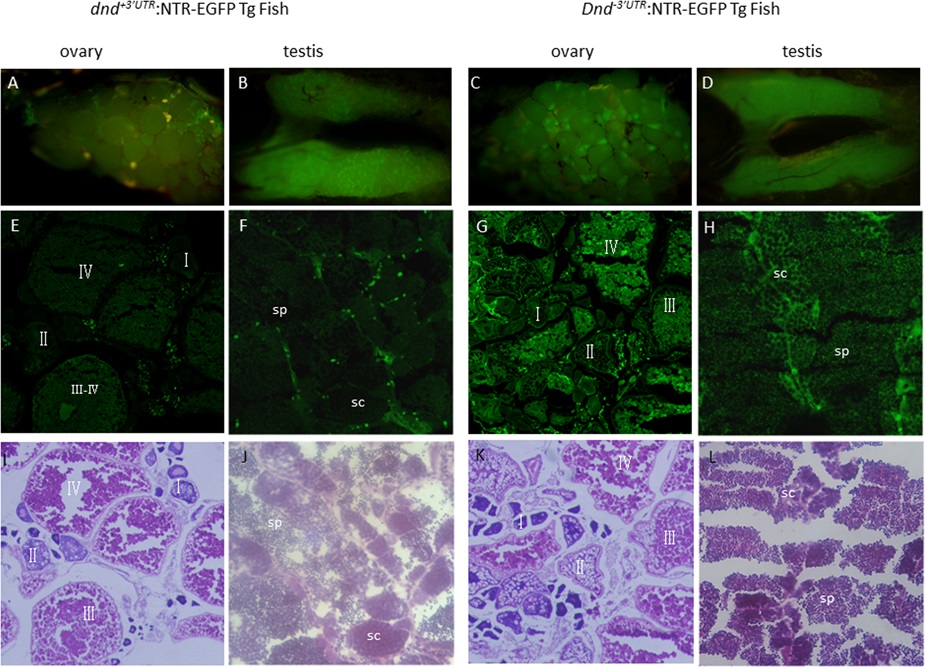
Fig. 3
Gonadal expression of the dnd+3’UTR:NTR-EGFP and dnd-3’UTR:NTR-EGFP transgene in adult zebrafish.
Anatomical views of the gonadal expression of the NTR-EGFP transgene are presented in A-D; Views of NTR-EGFP trangene expression and histological features with HE staining in the cyosectioned gonadal tissue are presented in E-H and I-L; A and B) Expression of the transgene in zebrafish ovary (A) and testis (B) in dnd+3’UTR:NTR-EGFP transgenic fish at 100 dpf; C and D) Expression of the transgene in zebrafish ovary (C) and testis (D) in dnd-3’UTR:NTR-EGFP transgenic fish at 100 dpf; E and F) NTR-EGFP transgene expression in the cryosections of the ovary (E) and testis (F) of the F1 progenies of dnd+3’UTR:NTR-EGFP transgenic fish at 100 dpf; G and H) NTR-EGFP transgene expression in the cryosections of the ovary (G) and testis (H) of the F1 progenies of dnd-3’UTR:NTR-EGFP transgenic fish at 100 dpf; I-J) histological features with HE staining for the I-J) shows the HE staining for the cryosections of the ovary (I) and testis (J) of the F1 progenies of dnd+3’UTR:NTR-EGFP transgenic fish at 100 dpf; K-L) shows the HE staining for the cryosections of the ovary (K) and testis (L) of the F1 progenies of dnd-3’UTR:NTR-EGFP transgenic fish at 100 dpf; Fluorescence was observed in the oocytes at all stages (E, G, I, K) and in spermatocytes(sc) and sperm(sp) (F, H, J, L).