Image
Figure Caption
Fig. 5, S1
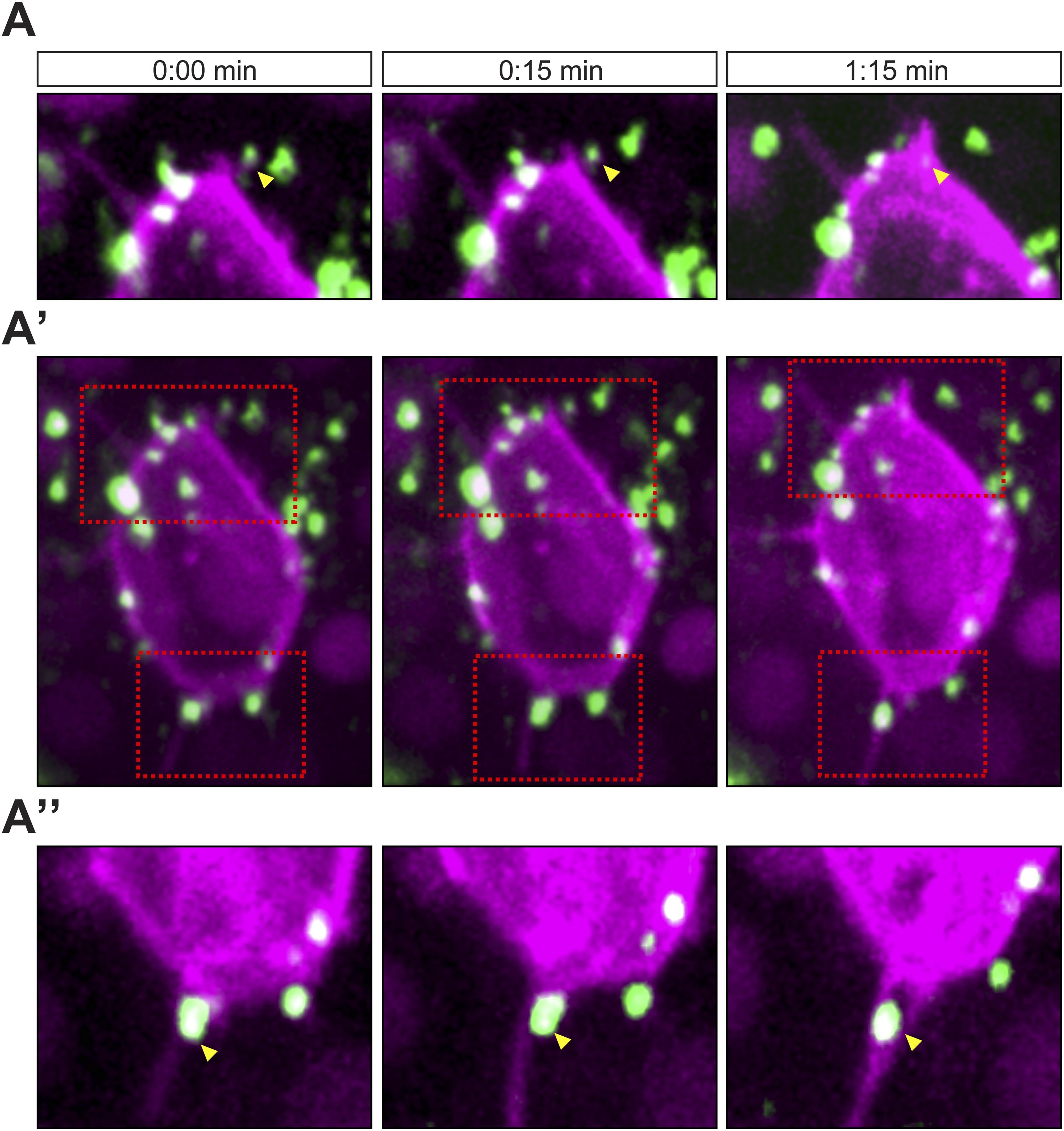
Cscl12a interaction with filopodia at the front and back of the cell
(A–A′′) Cxcl12a (Venus-tagged, green) interacts with filopodia of a PGC (mCherry-F, magenta). (A′) An optical section of a PGC (a Z-projection of 4 1-µm-slices) with Cxcl12a (green) interaction with filopodia (magenta) observed over 1:15 min. (A and A′′) Magnified insets marked in A′ as red squares. The yellow arrowheads point at Cxcl12a spots along filopodia.
Acknowledgments
This image is the copyrighted work of the attributed author or publisher, and
ZFIN has permission only to display this image to its users.
Additional permissions should be obtained from the applicable author or publisher of the image.
Full text @ Elife

