
|
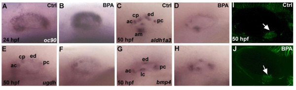
Fig. 3
Expression of markers of inner ear development in zebrafish embryos are affected by BPA treatment. (A,B)oc90 a gene require for otolith formation in zebrafish is up-regulated under BPA treatment at 24 hpf (n=30). In situ for control and BPA treated embryos were performed in the same tube with the tip of the tail removed for the control embryos. (C,D)aldh1a3 is detected in the anterior cristae (ac), the cranial epithelial projection (cp), the endolymphatic duct (ed), the posterior cristae (pc) and the anterior macula (am) in wild type zebrafish embryo at 50 hpf (M), its expression is reduced in ac, cp and am and is absent in ed and pc in BPA treated embryos from 6 hpf onward (D). (E,F) Expression of ugdh at 50 hpf in control otic vesicle (E) is detected in the ac, cp, ed and pc. In BPA treated embryos from 6 hpf onwards (D), ugdh expression is severely reduced and remains solely detected in the ac and the cp. (G,H) Expression of bmp4 in control embryos at at 50 hpf (G) in the anterior (ac) lateral (lc) in the posterior cristae (pc) and the endolymphatic duct (ed) remains unaffected after BPA treatment (H). (I,J) Confocal microscopy of acetulated tubulin antibody staining showing the presence of the ciliated macula (white arrow) in control embryos (I) and in BPA treated embryos (white arrow in J).