
Fig. 3
- ID
- ZDB-IMAGE-150520-11
- Publication
- Ju et al., 2015 - Oncogenic KRAS promotes malignant brain tumors in zebrafish
- All Figures
- Figures for Ju et al., 2015
|
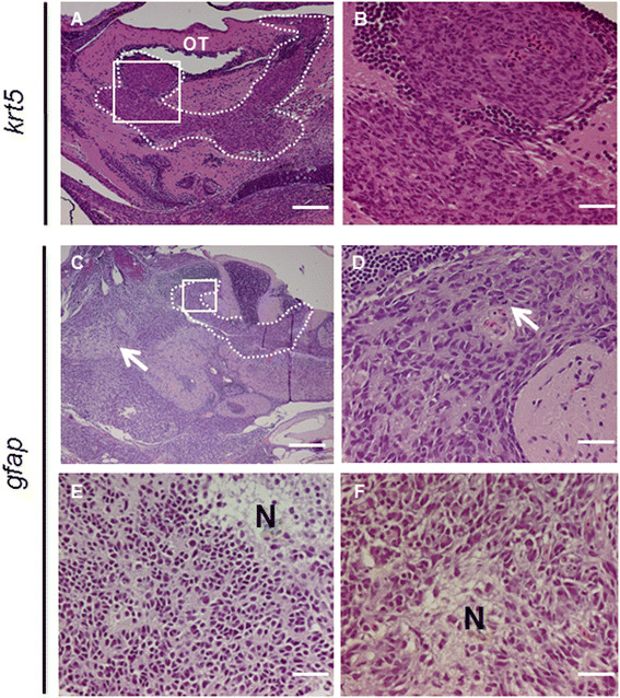
Fig. 3
Histological and immunohistological analyses of tumors fromkrt5andgfaptransgenic fish. (A) H&E staining of a sagittal section from a 6-month-old krt5-derived tumor showing tumor cells originating from and infiltrating the VZ of the optic tectum (OT), as highlighted by the dotted line. (B) Enlarged view of the white frame in (A) demonstrating infiltrating malignant cells with moderate pleomorphism, typical of high-grade astrocytoma. (C) A 6-month-old gfap-transgenic fish brain showing tumor development in both the VZ (broken line) and the brain parenchyma (arrow). gfap:KRASG12V-derived brain tumors exhibited phenotypes consistent with malignant glioma including frequent mitotic figures (D) and focal necrosis (E, F). N, necrosis. Scale bars, 100 µm for A; 20 µm for B, D, E and F; 40 µm for C.