
Fig. S3
- ID
- ZDB-IMAGE-150506-8
- Publication
- Ablain et al., 2015 - A CRISPR/Cas9 Vector System for Tissue-Specific Gene Disruption in Zebrafish
- All Figures
- Figures for Ablain et al., 2015
|
Fig. S3
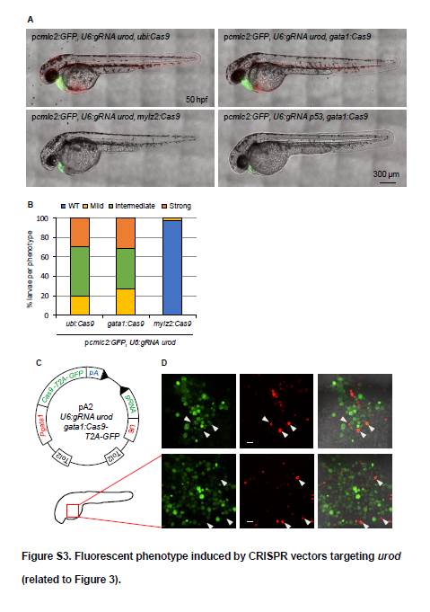
Fluorescent phenotype induced by CRISPR vectors targeting urod (related to Figure 3).
(A) Confocal images of 50 hpf embryos injected with Tol2 mRNA and pcmlc2:GFP, U6:gRNA urod vectors expressing Cas9 under the control of the indicated promoters.
(B) Quantification of the fluorescent phenotype presented in Figure 3A. WT: no fluorescent blood cell. Mild: <10 fluorescent blood cells. Intermediate: <50 fluorescent blood cells. Strong: >50 fluorescent blood cells. Ubi:Cas9 (n=61), gata1:Cas9 (n=51), mylz2:Cas9 (n=43).
(C) Schematic representation of a variant tissue-specific CRISPR vector. Gata1 promoter (Pgata1) drives both Cas9 and GFP expression through a self-cleaving T2A peptide. Any gRNA of interest can be produced ubiquitously off the U6 promoter. Tol2 indicates transposition sites for the Tol2 transposase. pA: SV40 polyA sequence.
(D) Confocal images of the yolk region of 30 hpf embryos injected with Tol2 mRNA and the pA2, U6:gRNA urod, gata1:Cas9-T2A-GFP vector. Arrows point to cells that are both green and red. Scale bar: 20 μm. Note the small size of red-only cells.
Reprinted from Developmental Cell, 32(6), Ablain, J., Durand, E.M., Yang, S., Zhou, Y., Zon, L.I., A CRISPR/Cas9 Vector System for Tissue-Specific Gene Disruption in Zebrafish, 756-64, Copyright (2015) with permission from Elsevier. Full text @ Dev. Cell