
Fig. S3
- ID
- ZDB-IMAGE-150506-55
- Publication
- Chou et al., 2014 - The Hemodynamically-Regulated Vascular Microenvironment Promotes Migration of the Steroidogenic Tissue during Its Interaction with Chromaffin Cells in the Zebrafish Embryo
- All Figures
- Figures for Chou et al., 2014
|
Fig. S3
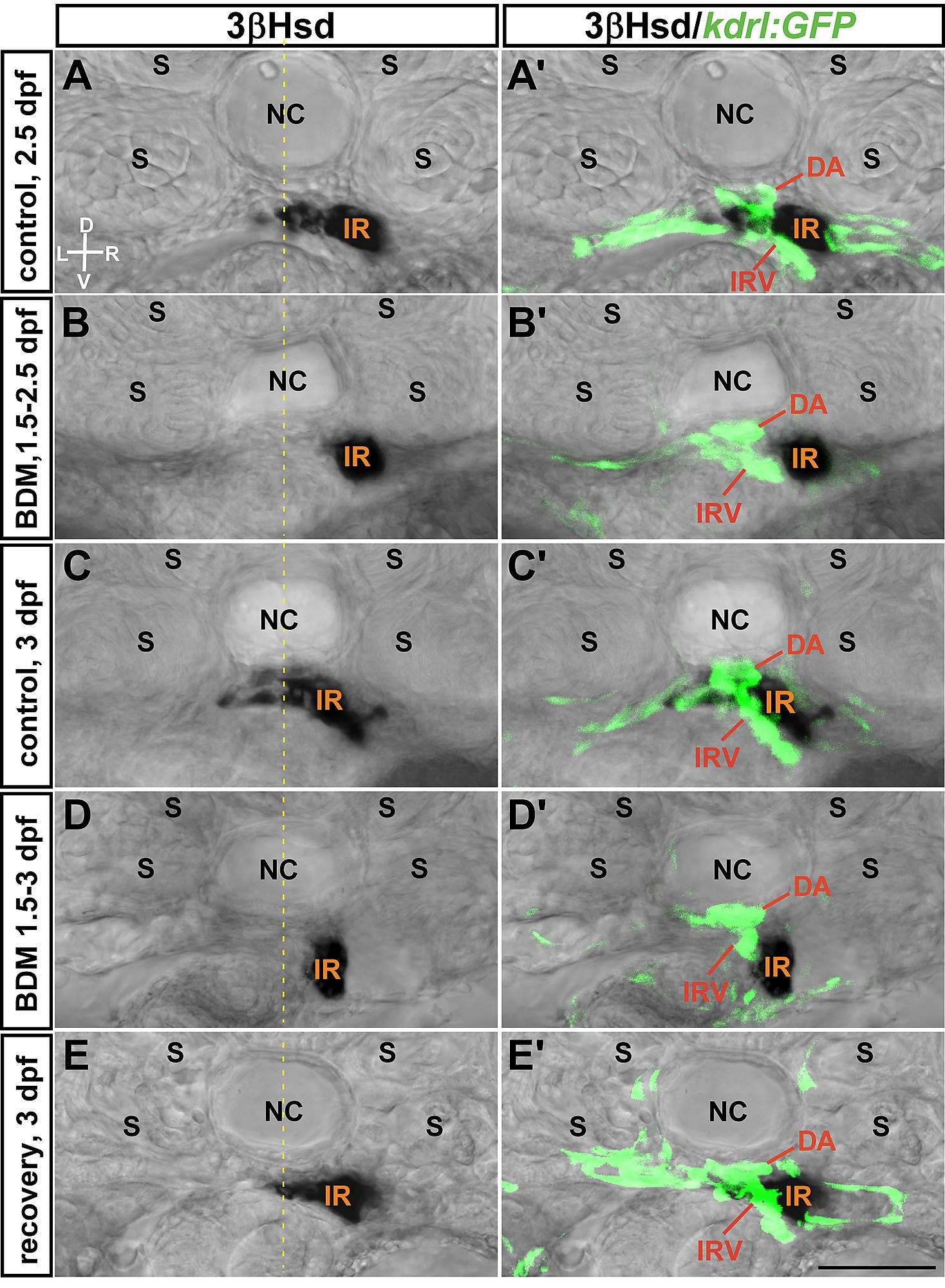
Effects of blood flow inhibition on IRV growth was reversible following the removal of 2,3-BDM.
The IRV in the control Tg(kdrl: GFP)s843 embryo continued to extend from 2.5 dpf (A, A′) to 3 dpf (C, C′), while the IRV growth was repressed by 2,3-BDM treatment at 6 mM from 1.5 to 2.5 dpf (B, B′) or 3 dpf (D, D′). Extension of IRV was recovered at 3 dpf as 2,3-BDM was washed out at 2.5 dpf (E, E′). The interrenal tissue (IR) was detected by 3β-Hsd acitivity assay. D, dorsal; V, ventral; L, left; R, right. Broken yellow lines indicate position of the midline. Abbreviations: notochord (NC), somite (S). Scale bar, 50 µm.