
IMAGE
Fig. 4
- ID
- ZDB-IMAGE-150430-9
- Publication
- Wen et al., 2013 - Synchronous and asynchronous modes of synaptic transmission utilize different calcium sources
- All Figures
- Figures for Wen et al., 2013
Image
|
Figure Caption
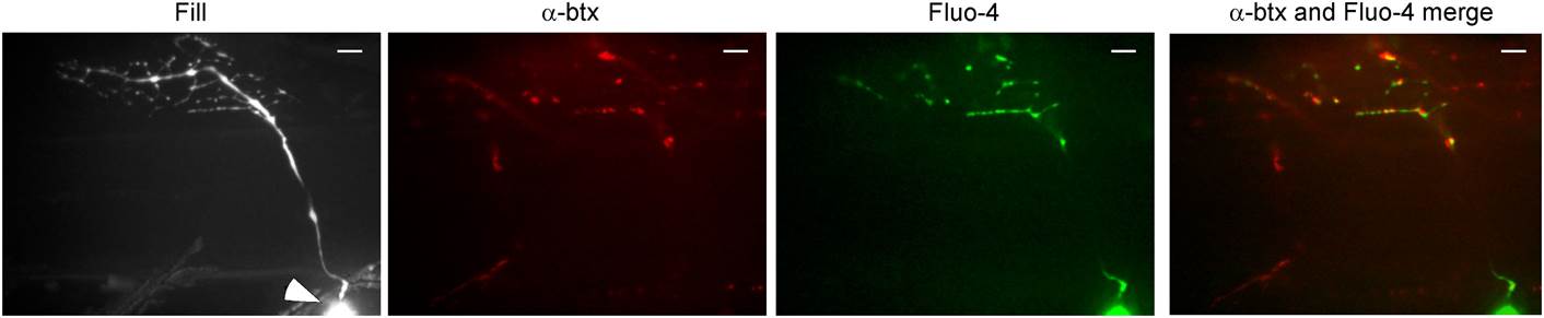
Fig. 4
Stimulus-driven calcium signals in CaP motor neuron terminals occurred at synaptic boutons.
The fill corresponds to a maximal intensity projection image of the motor neuron filled with Alexa Fluor 647. An arrowhead indicates the soma. A single plane of focus in the filled neuron showing postsynaptic α-btx label, peak Fluo-4 calcium signal and merged α-btx and Fluo-4 signal. The scale bar corresponds to 10 µm.
Acknowledgments
This image is the copyrighted work of the attributed author or publisher, and
ZFIN has permission only to display this image to its users.
Additional permissions should be obtained from the applicable author or publisher of the image.
Full text @ Elife