
Fig. 5, S1
- ID
- ZDB-IMAGE-150430-49
- Publication
- Lush et al., 2014 - ErbB expressing Schwann cells control lateral line progenitor cells via non-cell-autonomous regulation of Wnt/beta-catenin
- All Figures
- Figures for Lush et al., 2014
|
Fig. 5, S1
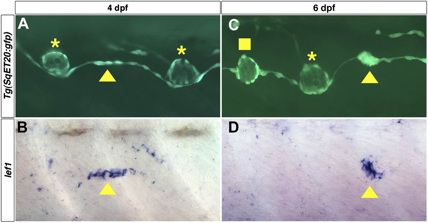
lef1 is expressed in clusters of interneuromast cells before they become intercalary neuromasts.
Lateral line cells expressing Tg(SqET20:gfp) were imaged at 4 or 6 dpf. Larvae were then fixed at processed for lef1 in situ hybridization and photographed at the same level. (A) At 4 dpf a small group of Tg(SqET20:gfp) positive interneuromast cells (arrowhead) can be seen between two primII deposited primary neuromasts (asterisk). This small cluster of interneuromast cells is positive for lef1 (B, arrowhead). (C) At 6 dpf a larger cluster of interneuromast cells is seen forming. Again this cluster of cells is lef1 positive (D, arrowhead). Interestingly, at 6 dpf one intercalary neuromast has formed (C, square), but it is not positive for lef1 (D). Suggesting lef1 is quickly decreased as neuromasts mature.