
Fig. 2
- ID
- ZDB-IMAGE-150428-8
- Publication
- Luz et al., 2013 - Fluorescently tagged Lin7c is a dynamic marker for polarity maturation in the zebrafish retinal epithelium
- All Figures
- Figures for Luz et al., 2013
|
Fig. 2
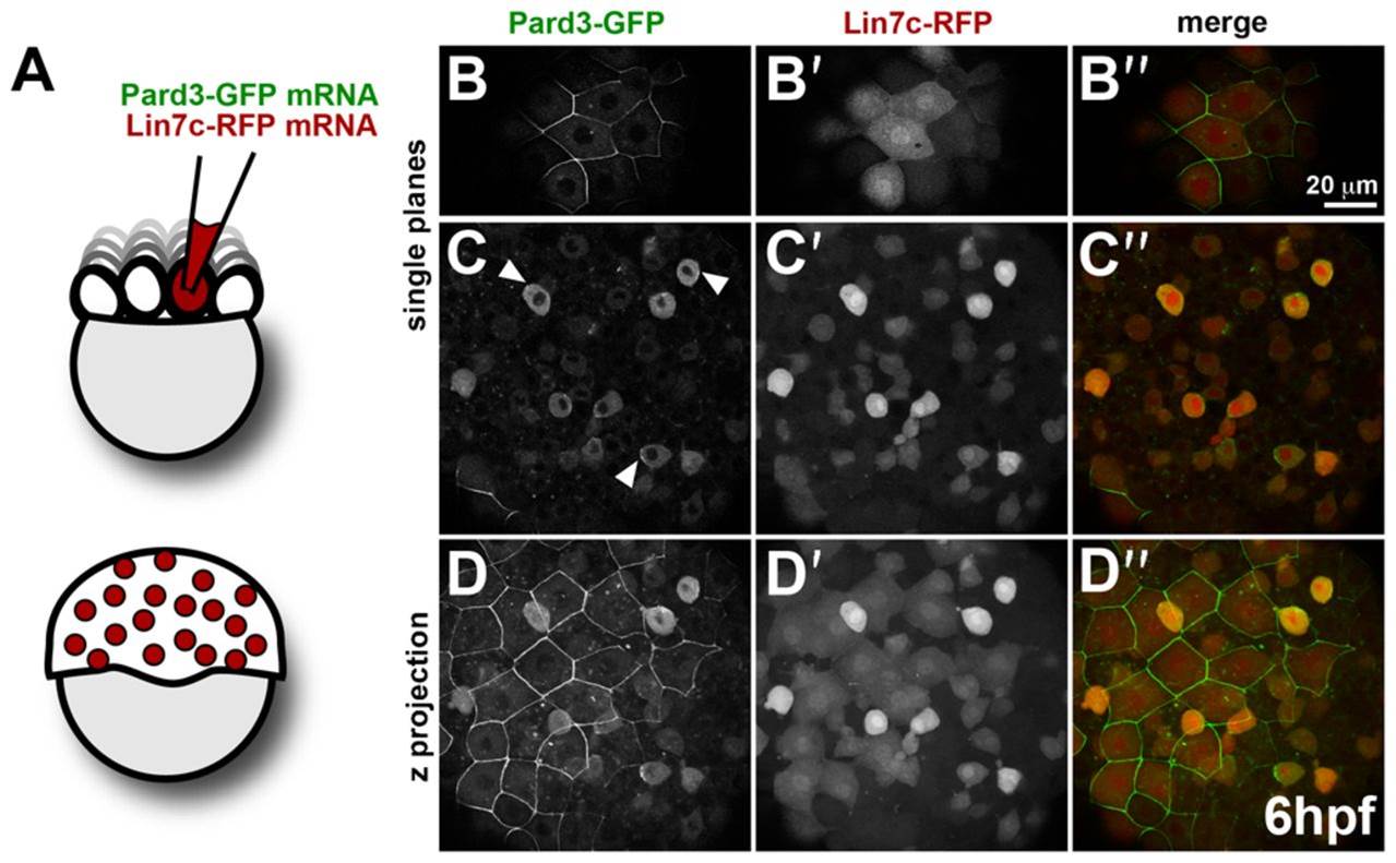
Comparison of Pard3-GFP and Lin7c-RFP subcellular localization reflects tissue-specific differences in epithelial polarity.
(A) Schematic representation of mRNA injection in 1/16–32 blastomeres for mosaic expression. (B–D′′) Mosaic co-expression of Par3-GFP and Lin7c-RFP in the gastrula (6 hpf). Par3-GFP is restricted to the membrane of the enveloping layer (EVL) cells (B), while it is mainly cytosolic in the deep cell layer (DEL), with some local accumulations at the cortex (arrowheads) (C). Lin7c-RFP is cytosolic and nuclear both in the EVL (B′) and DEL (C′). (D–D′′) Maximum projection of the embryo showing Pard3-GFP (D), Lin7c-RFP (D′) and the merge of both (D′′) in the EVL and DEL. Scale bar: 20 µm. Single confocal planes are shown, except for D–D′′.