
|
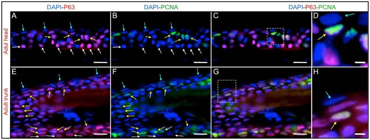
Fig. 6
PCNA and P63 immunofluorescence in the adult zebrafish epidermis.
(A–D) Head region. (E–H) Dorsal trunk area, in a region that contains only scales. (A and E) DAPI and P63 labeling. (B and F) DAPI and PCNA immunofluorescence. (C and G) Merged P63, PCNA and DAPI immunostainings. (D and H) Higher magnification images of the regions marked by squares in (C and G), respectively. Suprabasal P63-positive cells persist in adulthood and remain actively proliferating. Blue arrows mark cells from the external epidermal layer, yellow arrows label suprabasal cells at intermediate layers that are positive for PCNA and P63 expression, white arrows mark P63 positive cells from the basal layer. Bars are 20µm in length, except in (D, H and L) where they are 5µm in length.