
|
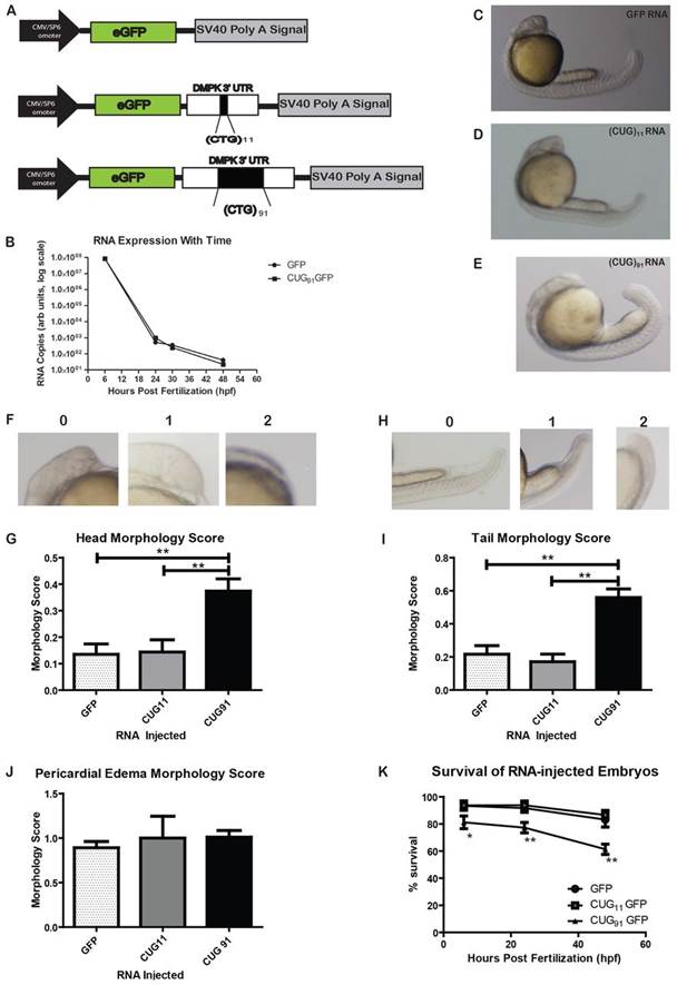
Fig. 1
Expanded CUG repeat RNA injection elicits morphological abnormalities in zebrafish embryos. (A) Schematic of constructs used to generate in vitro transcribed mRNA. (B) Injected RNA stability is similar for GFP and GFP(CUG)91 mRNAs, as assessed by qRT-PCR. Expression is normalized to actin mRNA from the same samples at all time-points; n=10 fish per group at each time point. (C-E) Representative embryos at 24 hpf injected with GFP mRNA (C), GFP(CUG)11 mRNA (D) or GFP(CUG)91 mRNA (E). (F) Abnormal head phenotypes observed in some GFP(CUG)91 mRNA-injected embryos at 24 hpf. 0, normal; 1, mild abnormalities; 2, severe abnormalities. (G) Blinded quantification of the abnormal head phenotypes across groups. (H) Abnormal tail and body shape phenotypes observed in some GFP(CUG)91 mRNA-injected embryos at 24 hpf. (I) Blinded quantification of the abnormal tail phenotypes across groups. (J) Blinded quantification of pericardial edema across groups. (K) GFP(CUG)91 mRNA-injected embryos have increased mortality at 24 and 48 hpf compared with GFP or GFP(CUG)11 mRNA-injected embryos. Graph shows survival of embryos injected with the indicated RNAs over 48 hours. Data from G, I, J and K represent n>200 embryos per group and at least five independent experiments. *P<0.05, **P<0.001. For morphological assessments, this represents the Dunn post-hoc multiple comparison test after confirmation of significant differences by the Kruskal-Wallis one-way ANOVA. For K, this represents a chi-squared test.