
Image
|
Figure Caption
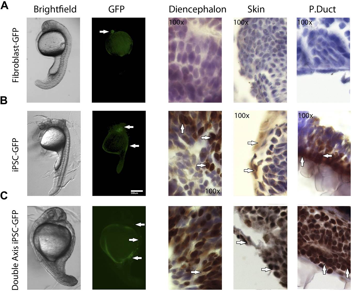
Fig. 6, S2
1 day old post fertilization zebrafish embryos.
(A) Embryos generated with control fibroblast cells exhibiting some localized flourescent cells. (B) Generated with iPSC-like GFP cells distributed in several parts of the embryo. (C) A double axis embryo, generated with iPSC-like GFP cells, showning one axis with high gfp flourescence, and none on the other. Explanation of histology sections is the same as in Figure 5.
Acknowledgments
This image is the copyrighted work of the attributed author or publisher, and
ZFIN has permission only to display this image to its users.
Additional permissions should be obtained from the applicable author or publisher of the image.
Full text @ Elife