
|
Fig. 2
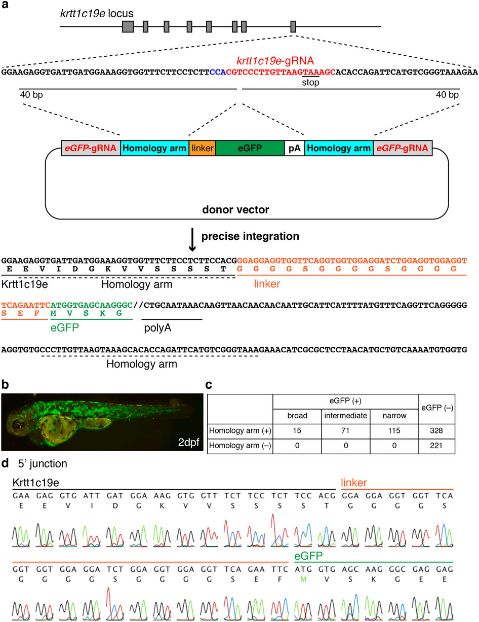
Precise integration of eGFP into the krtt1c19e locus.
(a) A schematic representation of the krtt1c19e locus and the donor vector consisting of eGFP-gRNA target sequences, homology arms, eGFP and polyA (pA) signal. The krtt1c19e-gRNA was designed to target the vicinity of the stop codon of the krtt1c19e gene. The upstream sequences of the krtt1c19e-gRNA target locus (krtt1c19e-gRNA sites in red, PAM sequence in blue) were inserted between the eGFP-gRNA target sequence and linker sequence on the donor vector, whereas the downstream sequences of the krtt1c19e-gRNA target locus were inserted between the polyA signal and eGFP-gRNA target sequence in the donor vector. When the donor vector, gRNAs and Cas9 mRNA were co-injected into 1–2-cell-stage embryos, the krtt1c19e gene and eGFP were connected in the same reading frame via the linker sequence by precise integration into the targeted genomic locus. (b) The injected embryo showed broad eGFP expression in the epidermis 2 days post-fertilisation (dpf). (c) The eGFP expression level was classified into three groups: broad, intermediate and narrow. Representatives of each expression level are shown in Supplementary Fig. S4. We observed no eGFP expression in embryos injected with the donor vector lacking homology arms. (d) Sequence analysis at the 5′ junction of the genome integrated with the donor vector harbouring homology arms.