
|
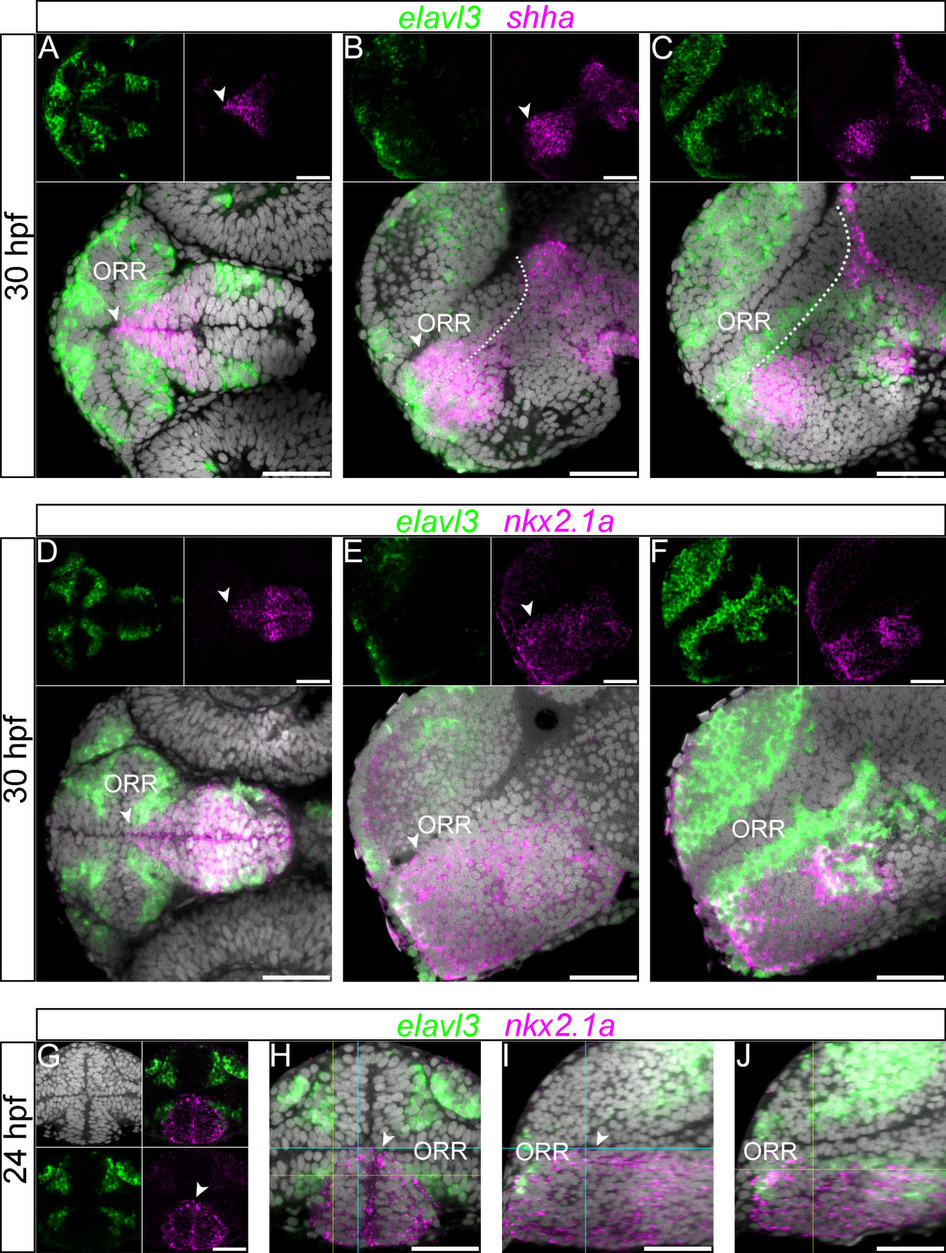
Fig. 5
Expression of nkx2.1a and shha in the hypothalamic region.
(A–C): 30hpf forebrain following elavl3 and shha in situ hybridization and DAPI staining (gray). The shha expression in the rostral edge of the hypothalamus displays a conical shape inserted into the ORR, which is visible in the ventral view (A). The expression reaches the ventricle at the most medial level (arrow heads of A and B), but not at a more lateral level (C).The currently proposed alar/basal limit is shown in dotted lines in the lateral sections (B and C), and it does not completely match the expression of shha in the anterior part of the forebrain. Scale bars = 50µm. (D–F): 30hpf forebrain following elavl3 and nkx2.1a in situ hybridization and DAPI staining (gray). The expression domain is also conical shaped in the ventral view (D), and it reaches the ventricle at the most medial level (arrow heads of D and E), but not at a more lateral level (F). (G–J): 24hpf forebrain following elavl3 and nkx2.1a in situ hybridization and DAPI staining (gray) illustrated in a single confocal plane of a frontal view (G and H) and two lateral views (I and J) reconstructed using Z-projections of the frontal images. Corresponding section levels of the two lateral views are indicated in blue (more medial) and yellow (more lateral) lines. The expression domain of nkx2.1a has a conical shape, visible in the frontal view (G and H). Its dorsal limit reaches the ventricle only in the section close to the midline (arrow heads in G–I) and not in the more lateral section (J).