
|
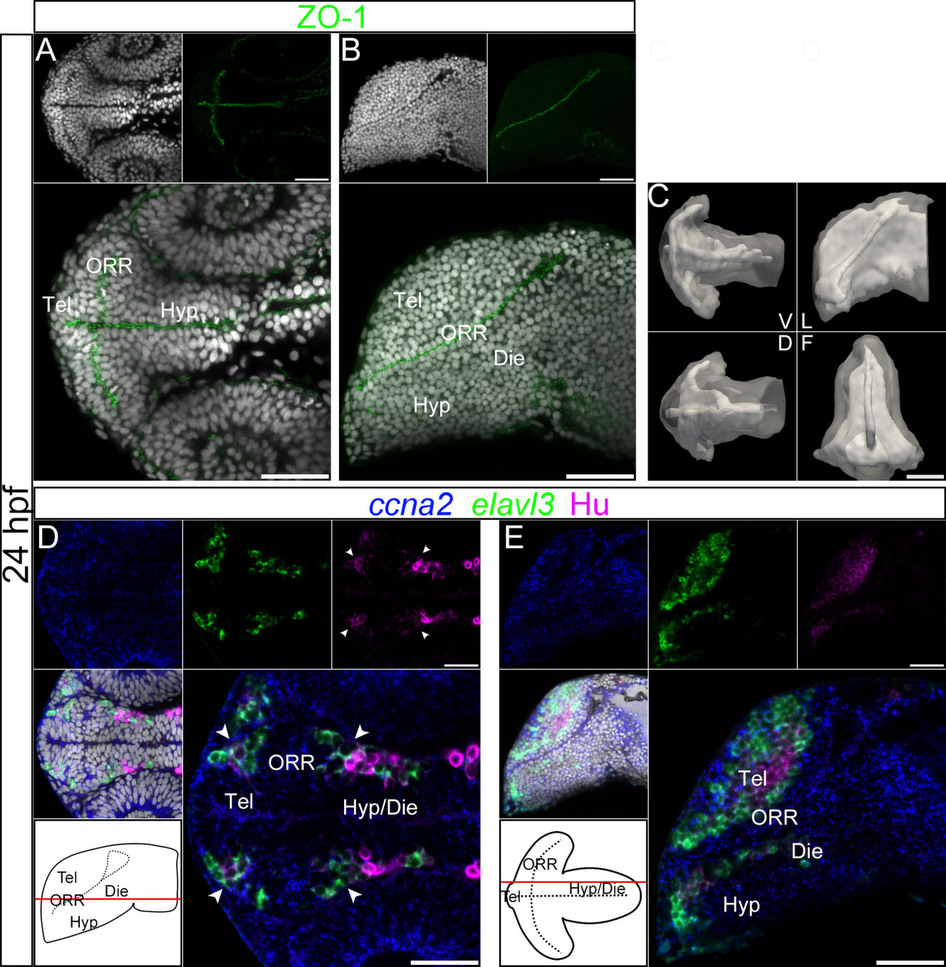
Fig. 3
Ventricular organization and the centrifugal gradient of neurogenesis at 24hpf.
(A–B): Single confocal plane of a 24hpf embryonic forebrain stained with DAPI (gray) and immunolabeled for ZO-1, in ventral (A) and lateral (B) views. diencephalon (Die); hypothalamus (Hyp); optic recess region (ORR); telencephalon (Tel). (C): Surface rendering of the shape of ventricle (white, in opacity), reconstructed from ZO-1 immunolabeling, overlapped on the shape of the brain (white, in transparency), reconstructed from DAPI staining. V = ventral, D = dorsal, L = lateral and F = frontal views. At 24hpf the organization of the ventricular system is considerably simpler than at 48hpf. The telencephalic ventricle rostral to the optic recess is not yet expanded (in contrast to 30hpf). (D–E): Single confocal plane of a 24hpf embryonic forebrain labeled for the neurogenic markers ccna2, elavl3 and HuC/D, in ventral (D) and lateral (E) views. Bottom left drawings show the level of corresponding optical plane. The ccna2 staining is widely distributed while elavl3 and especially HuC/D expressions are restricted to small peripheral territories. The arrow heads in D indicate Hu-expressing cells located at the boundaries of telencephalon/ORR and ORR/hypothalamus. Scale bars = 50µm.