
Image
|
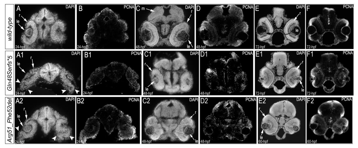
Figure Caption
Fig. S4
Immunostaining with PCNA (Proliferating Cell Nuclear Antigen) and DAPI in 24–72-hpf wild-type (A-F), mab21l2Q48Sfs*5 (A1-F1), and mab21l2R51_F52del (A2-F2) embryos.
Overlay images of the PCNA and DAPI immunostaining are shown in Fig. 6. The arrowheads in A1 and A2 indicate abnormal retinal folding;le, lens; m, midbrain; r, retina.
Acknowledgments
This image is the copyrighted work of the attributed author or publisher, and
ZFIN has permission only to display this image to its users.
Additional permissions should be obtained from the applicable author or publisher of the image.
Full text @ PLoS Genet.