
Fig. S9
- ID
- ZDB-IMAGE-150416-20
- Publication
- Cheng et al., 2015 - Nephron proximal tubule patterning and corpuscles of Stannius formation are regulated by the sim1a transcription factor and retinoic acid in zebrafish
- All Figures
- Figures for Cheng et al., 2015
|
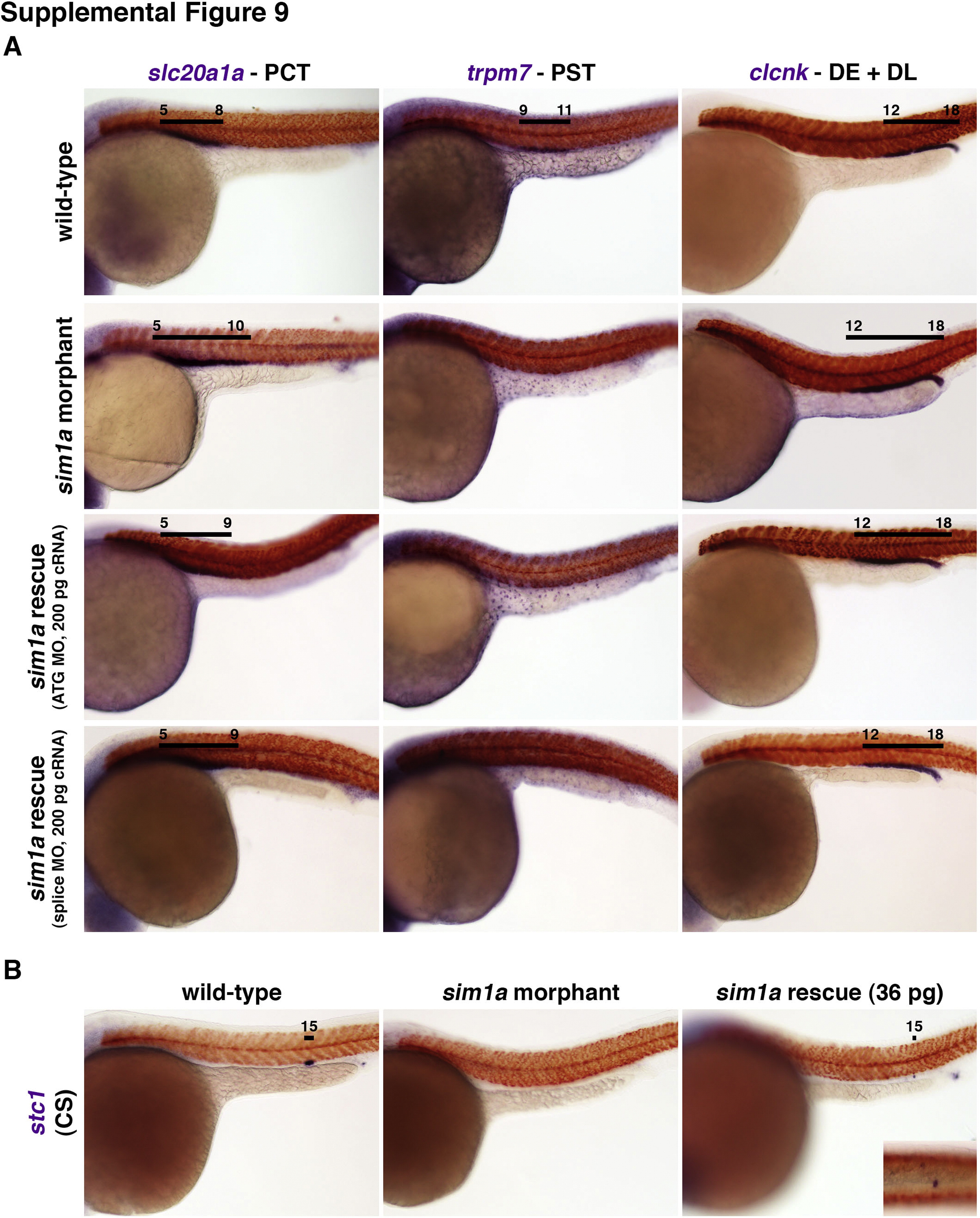
Fig. S9
sim1a overexpression is sufficient to partially rescue morpholino mediated sim1a deficiency. WISH analysis at 24 hpf of (A) wild-type control embryos, sim1a ATG morphants, sim1a ATG morphants co-injected with 200 pg of sim1a cRNA, or sim1a splice morphants co-injected with 200 pg of sim1a cRNA. Morphants (sim1a ATG or splice MO) that received sim1a cRNA showed a partial reduction in the PCT (slc20a1a) compared to sim1a morphants, but the PST (trpm7) was still abrogated. The distal tubule (clcnk) in these morphants was remained unchanged compared to the wild-type embryos. WISH analysis of (B) sim1a ATG morphants coinjected with 36 pg of sim1a cRNA showed a partial rescue of the CS (stc1) compared to sim1a ATG morphants. For all panels, renal marker expression (purple) and somite labeling by smyhc1 expression (red) are indicated. Black lines indicate segment domains in relation to corresponding somite numbers. Abbreviations: cRNA – capped RNA; CS – corpuscles of Stannius; DE – distal early; DL – distal late; hpf – hours post fertilization; PCT – proximal convoluted tubule; PST – proximal straight tubule; WISH – whole mount in situ hybridization.
Reprinted from Developmental Biology, 399(1), Cheng, C.N., Wingert, R.A., Nephron proximal tubule patterning and corpuscles of Stannius formation are regulated by the sim1a transcription factor and retinoic acid in zebrafish, 100-16, Copyright (2015) with permission from Elsevier. Full text @ Dev. Biol.