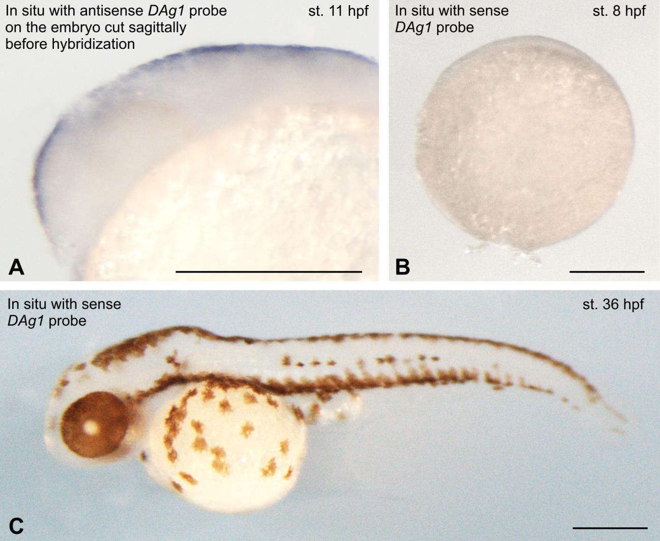
Fig. S1
Control in situ hybridization.
A. In situ with antisense DAg1 probe on the 11 hpf embryo cut sagittally before hybridization. DAg1 expression is seen only in the superficial layer of cells, but not in cells of deeper layers. This confirms that the in situ hybridization signal, which was observed primarily in the superficial cells of embryos hybridized in whole-mount, was not the result stipulated by a poor penetration of DAg1 probe into the depth of embryo. Anterior to the left, dorsal side up.
B and C. No signal was detected when embryos at 8 and 36 hpf were hybridized in whole mount with sense probe to DAg1. This result confirms specificity of the signal observed with antisense DAg1 probe. On C animal pole to the top. On C anterior to the left, dorsal side up. Scale bar everywhere is 200 µm

