
|
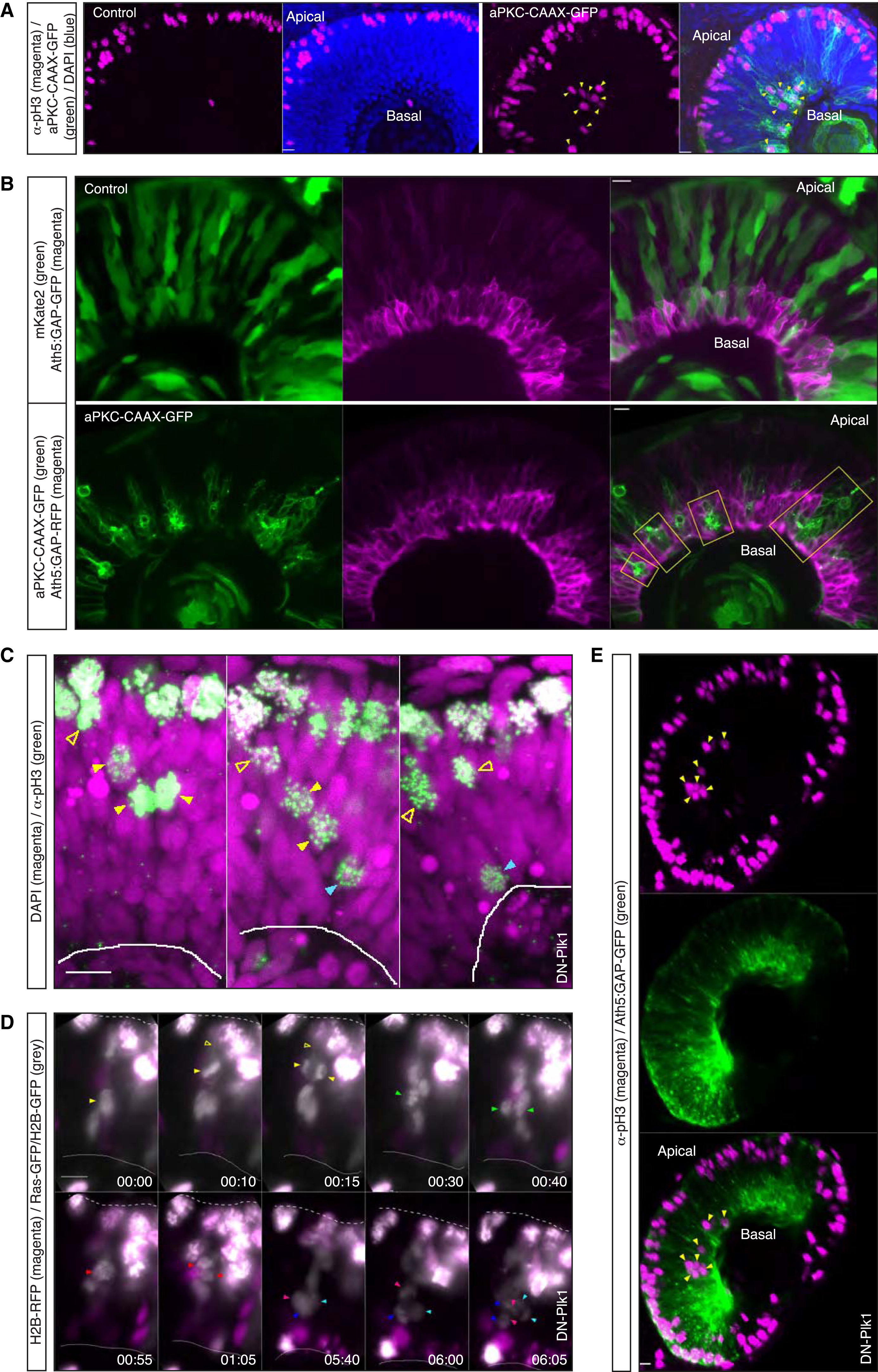
Fig. 7
Nonapical, Nonperpendicular Divisions Perturb Tissue Integrity and Early Neuronal Layering
(A) Confocal scans of a control embryo (left) and a HS-aPKC-CAAX-GFP injected embryo 24 hphs (right). Embryos were fixed at 50 hpf and stained with pH3 antibody. aPKC-CAAX expressing, mitotic cells can be observed at basal locations (arrows).
(B) Confocal scans of a control embryo injected with HS inducible cytosolic mKate2 (upper) and a HS-aPKC-CAAX-GFP injected embryo (lower) at 56 hpf/30 hphs. To visualize RGCs, the Tg(Ath5:GAP-RFP) line was used for aPKC-CAAX and Tg(Ath5:GAP-GFP) line for cytosolic mKate2 injection (RGCs, magenta). Control injected cells (green, upper) contribute to the intact RGC layer. In aPKC-CAAX expressing embryos the RGC layer features holes, filled by aPKC-CAAX (green) expressing cells (yellow boxes). See also Figure S4.
(C) Confocal scans of DN-Plk1 injected embryos stained with pH3 antibody (green) to visualize mitotic cells. DAPI is shown in magenta. DN-Plk1 expressing cells, arrested in mitosis, form an apical barrier. As a result, pH3-positive nuclei can be observed away from the apical surface (arrows: yellow open, subapical pH3; yellow filled, pH3 in the middle of the NE; cyan, basal pH3). HS was performed 11 hr prior to fixation.
(D) Time-lapse of cells in the embryo expressing DN-Plk1. DN-Plk1-positive cells coexpress H2B-RFP (magenta). To follow cell dynamics of nonmanipulated cells, H2B-GFP and Ras-GFP RNAs were injected mosaically (gray). Multiple non-DN-Plk1 cells can be observed dividing nonapically (arrows). Initially cells divide close to the apical barrier and maintain their apical process (yellow open arrows). Later in development, divisions occur at more basal locations (green, red, and blue arrows). HS was performed 11 hr prior to time-lapse. Time is in hr:min. The frames are from Movie S8. See also Figure S4.
(E) Confocal scans of Tg(Ath5:GAP-GFP) (green) embryos 53 hpf injected with DN-Plk1 and stained with pH3 antibody (magenta). pH3-positive nuclei can be observed at very basal locations (arrows). The RGC layer is disturbed in the central region of the retina, where the pH3-positive cells reside. HS was performed 29 hr prior to fixation. See also Figure S4.
Scale bars represent 10 µm. The dotted line represents the apical surface, and the solid line represents the basal side.
Reprinted from Developmental Cell, 32(2), Strzyz, P.J., Lee, H.O., Sidhaye, J., Weber, I.P., Leung, L.C., Norden, C., Interkinetic Nuclear Migration Is Centrosome Independent and Ensures Apical Cell Division to Maintain Tissue Integrity, 203-19, Copyright (2015) with permission from Elsevier. Full text @ Dev. Cell