
|
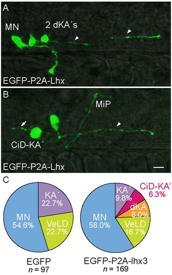
Fig. 6
Forced expression of Lhx3 in KA2s promotes descending and CiD axon trajectories. (A,B) Lateral views of 28hpf s1020t embryos. Labeled pMN derivatives express EGFP-P2A-Lhx3. (A) Pair of aberrant ventromedially located KA2 neurons with descending axons (dKA2, arrowheads). A rostral primary (RoP) MN projecting to lateral muscle is also labeled. (B) Aberrant ventromedially located KA2 neuron with an axon that initially extends laterally before turning caudally and descending within the spinal cord (CiD-KA2, arrowhead). An ascending axon collateral (arrow) sprouts immediately adjacent to the intraspinal portion of a labeled MiP MN. (C) Relative proportions of labeled pMN-derived neurons. Two new IN classes appear in UAS:EGFP-P2A-lhx3-injected embryos; KA2s are under-represented relative to control embryos (P=1.6×10⁵). The percentage of labeled cells is listed for each condition. Scale bar: 20µm.