
Fig. 6
- ID
- ZDB-IMAGE-150331-23
- Publication
- Shao et al., 2009 - Down syndrome critical region protein 5 regulates membrane localization of Wnt receptors, Dishevelled stability and convergent extension in vertebrate embryos
- All Figures
- Figures for Shao et al., 2009
|
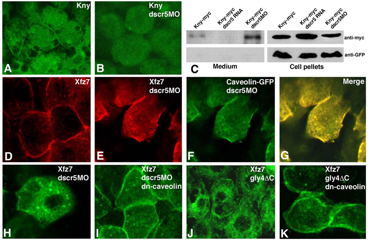
Fig. 6
Disruption of the membrane localization of Knypek and Frizzled 7 receptors in dscr5 morphants. (A) Localization of Kny-Myc at the cell surface and in the cytoplasm. (B) Kny-Myc distribution in dscr5-knockdown cells. (C) Western blot analysis of Kny-Myc levels in the culture medium (left) and cell pellets (right) of dscr5 RNA- or dscr5MO-injected cells. GFP protein was used as a loading control in the cell pellets and to monitor the integrity of the cultured cells. (D,E) XFz7-Myc localization revealed by Cy3-conjugated secondary antibody. (D) Localization of XFz7-Myc to the cell membrane of control cells. (E) XFz7-Myc distribution in dscr5-knockdown cells. (F) Caveolin-GFP localization in dscr5-knockdown cells. (G) Merge of E and F showing the colocalization between XFz7-Myc and Caveolin-GFP. (H-K) Rescue of XFz7-Myc membrane localization by dn-caveolin in cells injected with dscr5MO (I) or gly4ΔC RNA (K).