
|
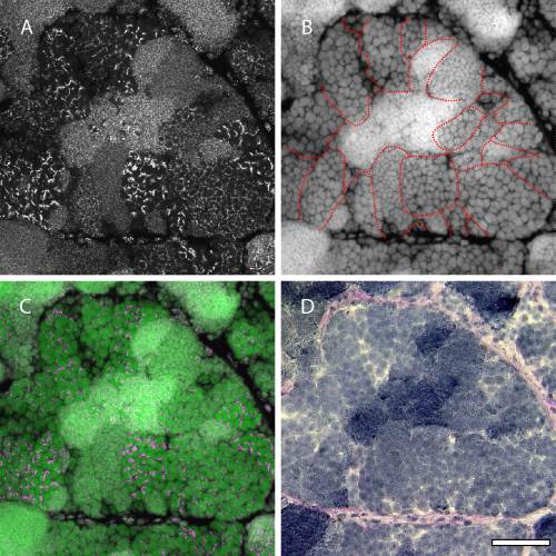
Fig. 2
Overall image of FISH signals using a telomere probe in zebrafish testis. A–C: FISH using a telomere probe in cryosections of a sexually mature testis. Panel A shows FISH signals, panel B shows nuclear staining with TO-PRO-3, and panel C shows a merged image of panels A and B. D: Same area viewed in panels A–C stained with periodic acid–Schiff (PAS) and hematoxylin. Zebrafish germ cells develop synchronously within each cyst (outlined in panel B). These cysts are formed by Sertoli cells in the tubular basement membrane, which is stained by PAS. Telomeres in the germ cells exhibit different patterns in each cyst. Scale bar = 50 µm.