
|
Fig. 7
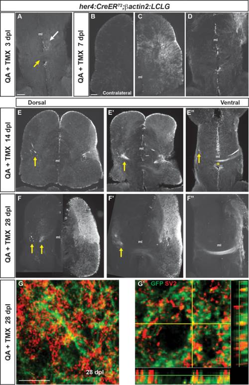
Neurons generated after QA-induced brain injury progressively extend processes to reestablish neuronal connections. (A) In the ventral telencephalon of Tg(her4:creERT2;β-actin2:LCLG) fish at 3 dpl, labeled progenitors began to extend GFP+ processes into the ipsilesional parenchyma (white arrow), but GFP expression was absent from the anterior commissure (yellow arrow) and contralateral hemisphere. (B–D) At 7 days after QA injection, minimal GFP expression was seen in the contralesional telencephalic hemisphere (B). In contrast, the ipsilesional hemisphere exhibited many GFP+ cells in the VZ and periventricular area that extended processes toward the injury site (C). At the level of the anterior commissure, GFP+ cells and processes were apparent but restricted to the ipsilesional hemisphere at 7 dpl (D). (E–E′′) Fasciculated bundles of GFP+ processes appeared by 14 dpl in the ipsilesional hemisphere and were also seen contralaterally (arrows) at dorsal levels (E, E2). More ventrally, GFP+ fibers appeared in the contralesional hemisphere (arrow in E3) and a GFP+ fiber bundle crossed at the level of the anterior commissure (E′′, asterisk). (F–F′′) The GFP+ bundles persisted at 28 dpl. At more dorsal levels, GFP+ fascicles were seen in the parenchyma of the contralesional hemisphere (F,F′), and at ventral levels an extensive fiber bundle crossed the anterior commissure (F′′). (G–G′) Colabeling for GFP (green) and the synaptic marker SV2 (red) in the contralesional telencephalon at 28 dpl revealed the appearance of synaptic contacts made by GFP+ axons onto putative targets. G′ shows an orthogonal projection of GFP+ synaptic contacts colabeled for SV2 (red). Scale bars = 100 µm. Scale bar in A applies to D, F′, F′&prime& in B to C, E, F. ml = midline.