
|
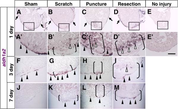
Fig. 4
Expression of aldh1a2 after various injuries. A–D, A′–D′, F–M: In situ hybridization of aldh1a2 at 1 day (A–D, A′–D′), 3 days (F–I), and 7 days (J–M) after various injuries. E: In situ hybridization of aldh1a2 without injury. A′–E′ show close up images of boxed areas in A–E. A, A′, F: Arrowheads and arrows point to evident signals in the epicardium at the apex and away from the apex, respectively. B–D, B′–D′, G–I, K–M: Arrowheads and arrows point to evident signals in the epicardium around injury sites and away from injury sites, respectively. Open arrowheads point to endocardial signals. Brackets indicate the injury sites. Scale bar = 50 µm.