
Image
|
Figure Caption
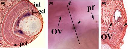
Fig. 3
rxfp3-2b expression territories outside the brain. (a) Counter-stained transverse sections of eye of larvae at 96 h postfertilization (hpf). (b) Whole-mount in situ hybridized larvae (96 hpf); particular of pharyngeal arch region. (c) Counter-stained transverse section of larvae at 96 hpf as indicated in (b). Black arrowhead indicates rxfp3-2b-expression cells in the thymus region. gcl, ganglion cell layer; inl, inner nuclear layer; OV, otic vesicle; pcl, photoreceptor cell layer; pf, pectoral fin.
Figure Data
Acknowledgments
This image is the copyrighted work of the attributed author or publisher, and
ZFIN has permission only to display this image to its users.
Additional permissions should be obtained from the applicable author or publisher of the image.
Full text @ Dev. Growth Diff.