
|
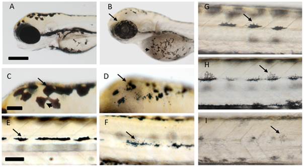
Fig. 3
Changes in melanocyte patterning and distribution following cigarette smoke condensate (CSC) exposure from 48–72 hpf.
A, C, E, G) DMSO Carrier control (CC). Changes in pigment distribution (arrows) within the melanphores of the eyes and yolksac (B), dorsal regions of the head (D) and the tail midline (F) following treatment with 50 µg/mL CSC prepared from 3R4F using the ISO smoking regime, are evident. Concentration-dependent change in melanocyte morphology and pigment level (arrows) following exposure to CSC prepared from the Canadian best seller (BSV) using the ISO smoking regime (H-6.1 µg/mL; I-50 µg/mL). Scale bars in A, C and E correspond to 280, 70 and 80 µm, respectively.