
|
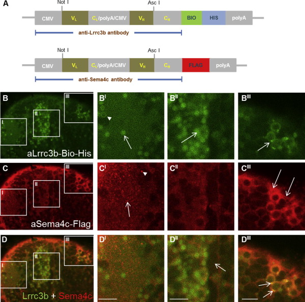
Fig. 4
The addition of different tags facilitates multiplex staining. (A) Schematics showing the addition of C-terminal Bio-6-His and FLAG tags to plasmids. The region encoding the specific antibodies are flanked by unique NotI and AscI sites facilitating antibody tag switching by subcloning. (B–D) The same 5 dpf embryo was simultaneously stained with both the biotin-tagged anti-Lrrc3b and FLAG-tagged anti-Sema4c antibodies. The staining of anti-Lrrc3b in green (B) and anti-Sema4c in red (C), together with the merged images (D) at the same site are shown. Staining patterns are described in the main text. Scale bars represent 50 µm.