
Fig. 1
- ID
- ZDB-IMAGE-150324-33
- Genes
- Publication
- Ceci et al., 2014 - Axon-Schwann cell interactions during peripheral nerve regeneration in zebrafish larvae
- All Figures
- Figures for Ceci et al., 2014
|
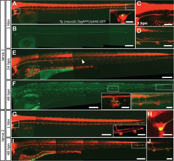
Fig. 1
Neuromast reinnervation after PLL nerve regeneration. Tg(neurod:TagRFP) fish that have a red labeled pLL nerve were injected at the one cell stage with the pE46:GFP DNA construct and were selected if they displayed green fluorescence in a single sensory neuron in the pLL ganglion (inset in A). The innervation pattern of the single sensory neuron was recorded both before neurectomy and after regeneration of the axon. Two different examples of reorganization during pLL nerve regeneration are shown, referred to as larva 1 (A-F) and larva 2 (G-J). Larva 1 shows a pLL ganglion neuron that innervates the terminal neuromasts before injury (B, D). Neurectomy is carried out about 200 µm away from the ganglion severing all axons of the pLL nerve (C). Twenty-four hours post neurectomy (hpn), the pLL nerve has regenerated about half way down the body of the larva (E). At 48 hpn, the nerve has completely regenerated (F) altough the green-labeled axon now innervates a different neuromast, the L4 (small inset in F) and does not innervate its original targets, which are innervated by other neurons (large inset in F). Larva 2 shows innervation of the terminal neurmasts before injury (G, H) and, after neurectomy and regeneration, the same neuromasts are reinnervated (I, J). Scale: A, B, E, F, G, I: 200 µm; C: 100 µm; inset in A, D, larger inset in F, inset in G, H; small inset in F: 20 µm.