
Fig. 7
- ID
- ZDB-IMAGE-150319-8
- Genes
- Publication
- Takeuchi et al., 2015 - Establishment of Gal4 transgenic zebrafish lines for analysis of development of cerebellar neural circuitry
- All Figures
- Figures for Takeuchi et al., 2015
|
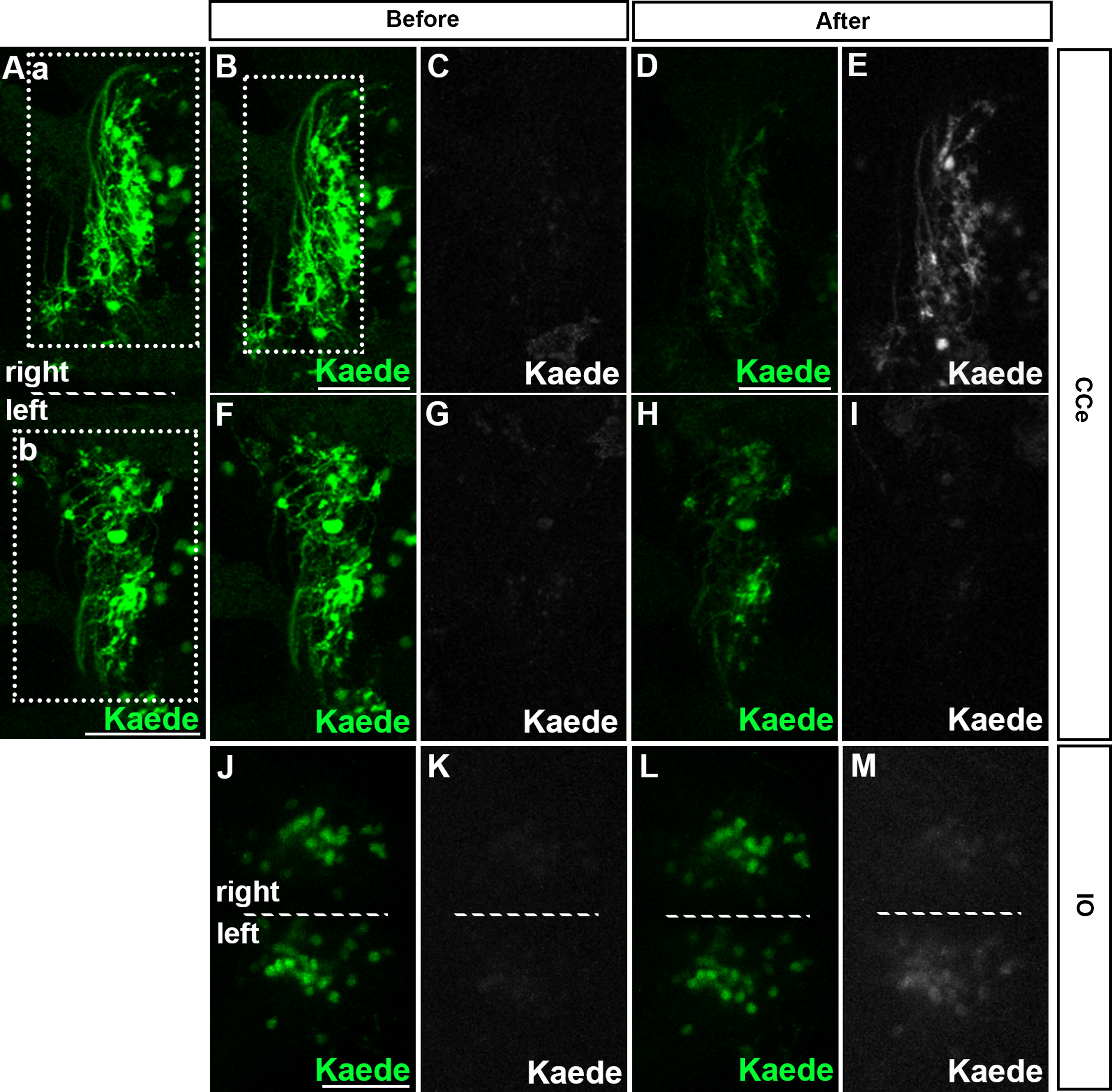
Fig. 7 Tracing with Kaede shows contralateral projections of CFs in zebrafish. A CF terminal region of the left side of 5-dpf hspGFFDMC28C; UAS:Kaede larvae was laser irradiated. Expression of Kaede and photoconverted Kaede were examined. Cerebellum (CCe, A–I) and IO (J–M) regions. Dorsal views with anterior to the left. The midlines are indicated by dotted lines in A, J–M. (B–E) High magnification views of the box a in A (right side of the cerebellum). (F–I) High magnification views of the box b in A (left side). The CF axon termini on the right side were irradiated. The irradiated area is marked by a dotted box in B. Expression of green (A, B, D, F, H) and photoconverted Kaede (white, C, E, G, I) was detected in the cerebellum (CCe) before (B, C, F, G) and 7 h after laser-mediated photoconversion (D, E, H, I). Expression of green (J, L) and photoconverted Kaede (K, M) was also detected in the IO region before (J, K) and 7 h after the photoconverstion (L, M). Similar contralateral projections were detected from twenty independent experiments. Scale bars: 50 µm in A; 50 µm in B (applied to B, C, F, G); 50 µm in D (applied to D, E, H, I); 40 µm in J (applied to J–M).
Reprinted from Developmental Biology, 397(1), Takeuchi, M., Matsuda, K., Yamaguchi, S., Asakawa, K., Miyasaka, N., Lal, P., Yoshihara, Y., Koga, A., Kawakami, K., Shimizu, T., Hibi, M., Establishment of Gal4 transgenic zebrafish lines for analysis of development of cerebellar neural circuitry, 1-17, Copyright (2015) with permission from Elsevier. Full text @ Dev. Biol.