
IMAGE
Fig. S1
- ID
- ZDB-IMAGE-150319-12
- Publication
- Takeuchi et al., 2015 - Establishment of Gal4 transgenic zebrafish lines for analysis of development of cerebellar neural circuitry
- All Figures
- Figures for Takeuchi et al., 2015
Image
|
Figure Caption
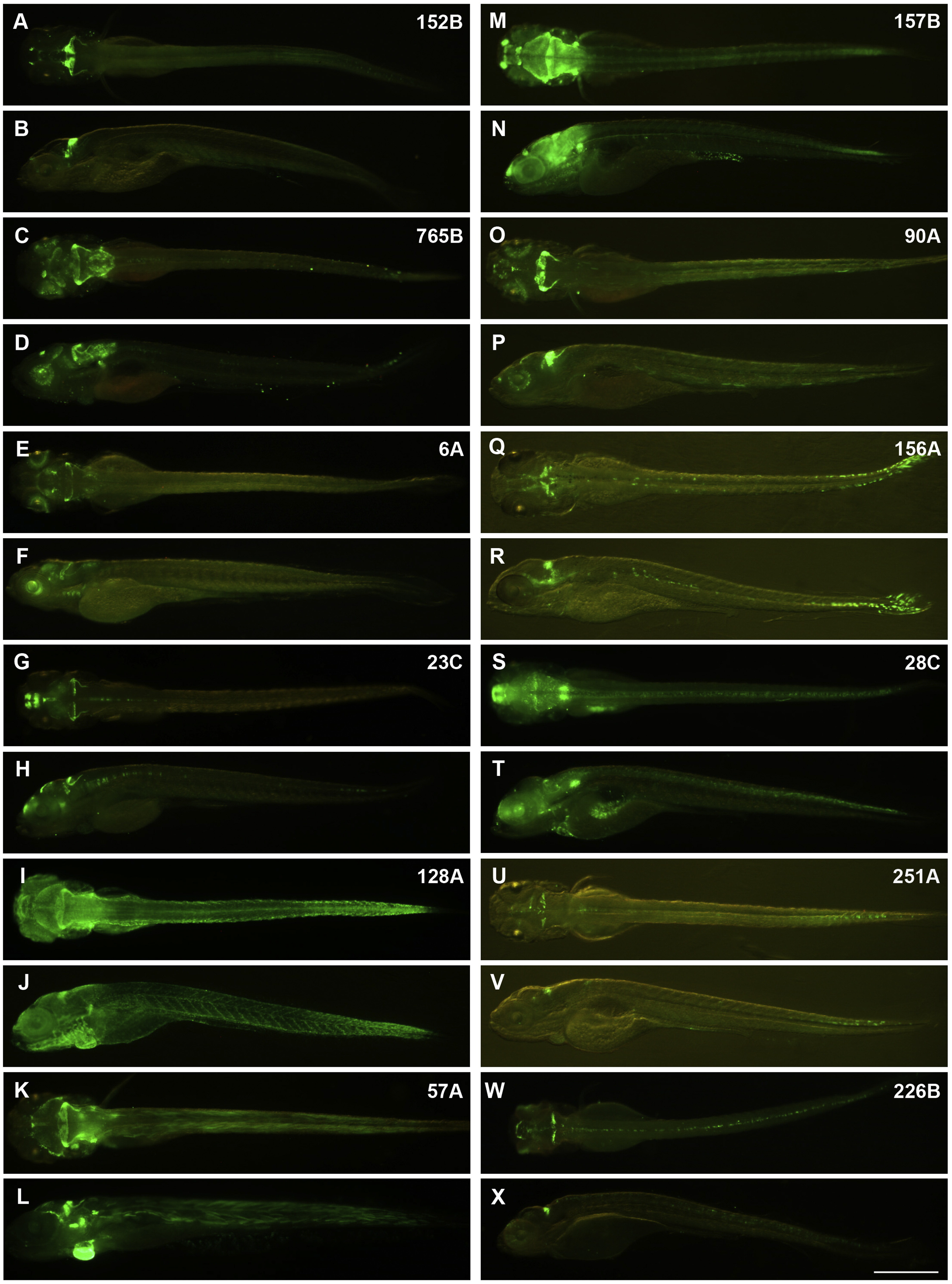
Fig. S1 Trap lines. Expression of GFP. 5-dpf larvae. (A, B) gSA2AzGFF152B; UAS:GFP. (C, D) gSAIzGFFM765B, UAS:GFP. (E, F) gSAG6A. (G, H) gSAIGFF23C; UAS:GFP. (I, J) SAGFF(LF)128A; UAS:EGFP. (K, L) hspGFF57A; UAS:GFP. (M, N) SAGFF(LF)157B; UAS:GFP. (O, P) hspGFFDMC90A; UAS:GFP. (Q, R) hspzGFFgDMC156A; UAS:GFP. (S, T) hspGFFDMC28C; UAS:GFP. (U, V) SAGFF(LF)251A; UAS:GFP. (W, X) SAGFF(LF)226B; UAS:GFP. Dorsal views with anterior to the left (A, C, E, G, I, K, M, O, Q, S, U, W). Lateral views with anterior to the left (B, D, F, H, J, L, N, P, R, T, V, X). Scale bar: 50 µm (applied to all panels).
Acknowledgments
This image is the copyrighted work of the attributed author or publisher, and
ZFIN has permission only to display this image to its users.
Additional permissions should be obtained from the applicable author or publisher of the image.
Reprinted from Developmental Biology, 397(1), Takeuchi, M., Matsuda, K., Yamaguchi, S., Asakawa, K., Miyasaka, N., Lal, P., Yoshihara, Y., Koga, A., Kawakami, K., Shimizu, T., Hibi, M., Establishment of Gal4 transgenic zebrafish lines for analysis of development of cerebellar neural circuitry, 1-17, Copyright (2015) with permission from Elsevier. Full text @ Dev. Biol.