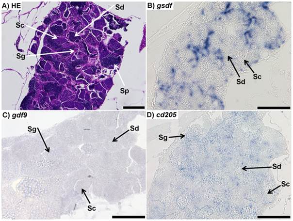
Fig. 2
Sections of testis from 7 month-old zebrafish.
A) Respresentative haematoxylin-eosin (HE) staining of the mature zebrafish testis; spermatogonia (Sg), spermatocytes (Sc), spermatids (Sd), and spermatozoa (Sp) could all be positively identified. B) in situ hybridization (ISH) of gsdf transcripts in the testis; staining shows strong signal throughout the testis, although it is unclear from which cell type it originates; no staining is visible in spermatocytes or spermatids. Spermatogonia and spermatozoa could not be identified. C) ISH of gdf9 transcripts in testis; faint signal was found throughout the testis. It is unclear which cell types the signal is localized to. D) ISH of cd205 transcripts in the testis; the signal is strongest in spermatogonia, while it is not visible in spermatocytes or spermatids. Spermatozoa could not be identified. All scalebars represent 100 µm.

