
Fig. 1
- ID
- ZDB-IMAGE-150313-32
- Publication
- Presslauer et al., 2014 - Induced Autoimmunity against Gonadal Proteins Affects Gonadal Development in Juvenile Zebrafish
- All Figures
- Figures for Presslauer et al., 2014
|
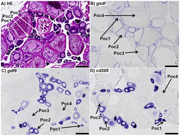
Fig. 1
Sections of ovaries from 7 month-old zebrafish.
A) Representative haematoxylin-eosin (HE) staining of the mature zebrafish ovary. B) in situ hybridization (ISH) of gsdf transcripts in the ovary; staining shows gsdf transcripts are localized in somatic cells surrounding primary oocytes. The staining is strongest in stage 2 primary oocytes (Poc2) and previtellogenic oocytes (Poc3). C) ISH of gdf9 transcripts in the ovary; strong signal is detected in stage 1 and 2 primary oocytes (Poc1 and Poc2). Weak signal is detected in previtellogenic oocytes (Poc3), while no signal was detected in vitellogenic oocytes (Poc4). D) ISH of cd205 transcripts in the ovary; strong signal is detected in stage 1 and 2 primary oocytes, with weak signal in previtellogenic oocytes, and no transcript detected in vitellogenic oocytes. All scalebars represent 200 µm.