
|
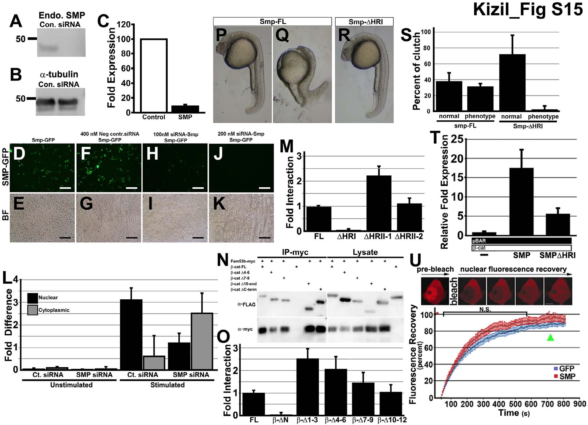
Fig. S15 Reduction of SMP protein by transfection of Smp siRNA, Related to Figure 4. (A) Western blot of immnoprecipitation pull down of β-catenin with different mutants of the Smp protein. (B) Western blot of antibody to the endogenous SMP protein in control siRNA- and SMP siRNA-transfected HEK293T cells. (C) Same blot samples probed with α-tubulin antibody. (D) Measurement of fold expression detected from Western blot of siRNA knockdown experiment. (E-L) Expression of GFP fused to the C-terminal of Smp. (E) Smp-GFP alone and (F) Bright field image of (E). (G) Smp-GFP in cells transfected with control siRNA. (H) bright field of (G). (I) SMP-GFP fluorescence in the cells transfected with 100 nM siRNA to Smp. (K) SMP-GFP in cells transfected with 200 nM of siRNA to Smp. (M) Lucifierase reporter assay for control β-catenin, Smp-FL and Smp delta homology region (I). (N) Fluorescence recovery curves after photobleaching mCherry-β-catenin in nucleus of cell transfected either with GFP or Smp. Scale bars equal 100 μm.