
|
Fig. S3
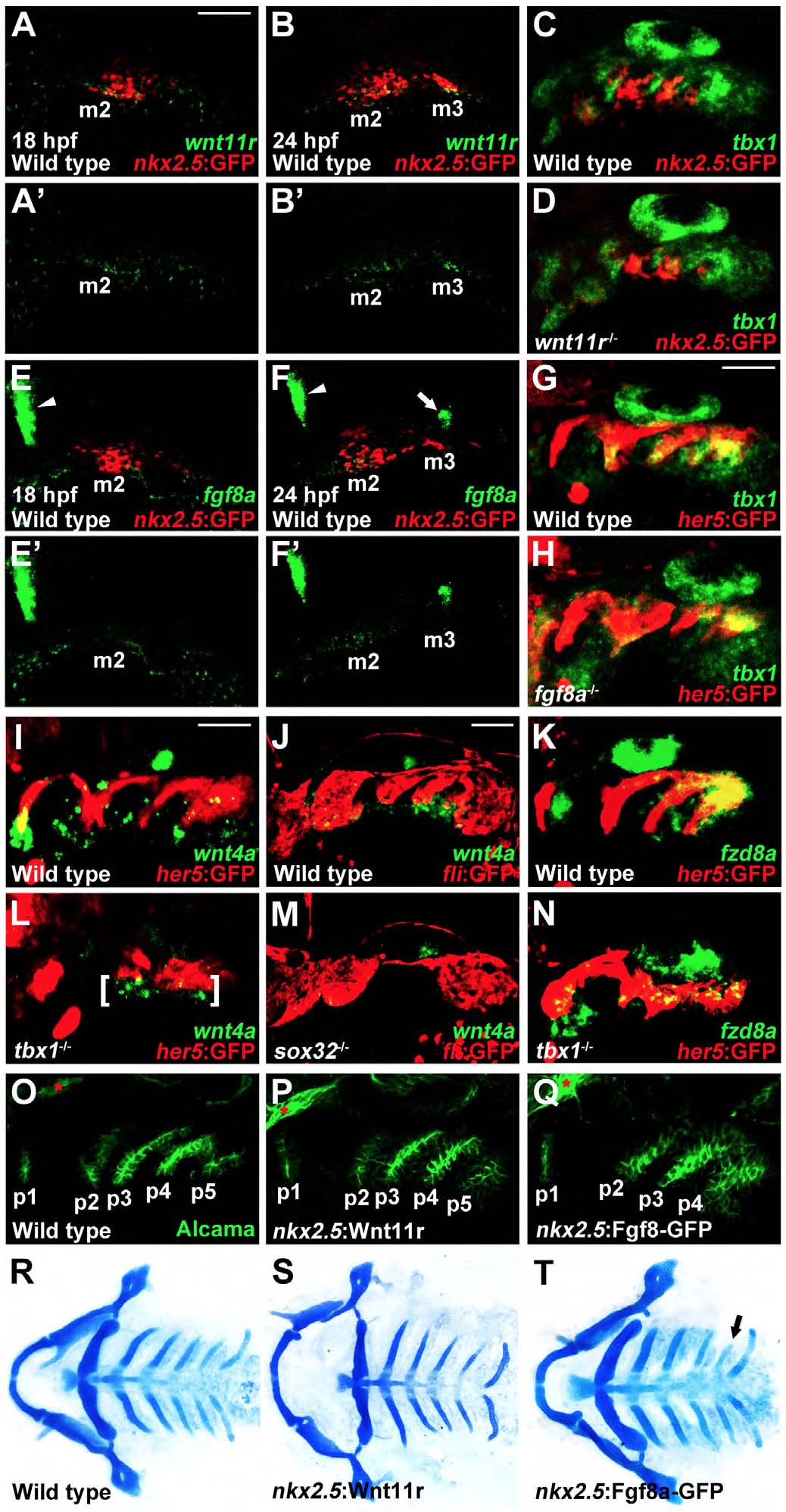
wnt4a and fzd8a expression in tbx1 mutants and analysis of nkx2.5:Wnt11r and nkx2.5:Fgf8a-GFP transgenes
(A-H) Fluorescent in situ hybridization for wnt11r, tbx1, or fgf8a mRNA (green). In red, GFP immunohistochemistry detects nkx2.5:GFP+ mesoderm (A-F) or her5:GFP+ endoderm (G,H). At 18 hpf, wnt11r is expressed in the second arch mesodermal core (m2) (A, n=17), and fgf8a is not yet expressed in the nkx2.5:GFP+ mesoderm (E, n=21). By 24 hpf, nkx2.5:GFP labels the third arch mesodermal core (m3) and wnt11r is expressed in both m2 and m3 (B, n=15) and fgf8a just in m2 (F, n=20). fgf8a mRNA expression is also observed at the midbrain-hindbrain boundary (arrowheads in E and F) and the anterior region of the otic vesicle (arrow in F). At 30 hpf, tbx1 expression is unaffected in 8/8 wnt11r mutants (D) or 11/11 fgf8a mutants (H) compared to wild-type siblings (C and G).
(I-N) Fluorescent in situ hybridization for wnt4a or fzd8a mRNA (green) at 30 hpf. In red, GFP immunohistochemistry detects her5:GFP+ endoderm (I,K,L,N) or fli1a:GFP+ neural-crest-derived ectomesenchyme (J,M). Compared to wild types (n=61), ectodermal wnt4a expression (brackets in L) persists but is disorganized in 18/18 tbx1 mutants. In contrast, 9/9 sox32-/- embryos displayed near complete loss of wnt4a ectodermal expression, with the lack of posterior arch segmentation indicative of the absence of pouch endoderm. Compared to normal expression in wild types (n=38), fzd8a expression within the her5:GFP+ endoderm is largely unaffected in 10/14 tbx1 mutants.
(O-Q) Alcama immunohistochemistry shows a series of five pouches at 34 hpf. Compared to wild types, we observed normal pouches in 49/49 nkx2.5:Wnt11r and 27/36 nkx2.5:Fgf8a-GFP embryos. In 9/36 nkx2.5:Fgf8a-GFP embryos, we observed delayed and/or disorganized formation of the last pouch. Sensory ganglia are indicated with red asterisks. Anterior is to the left and ventral is down. Scale bar represents 40 μm.
(R-T) Ventral whole-mount views of Alcian-stained facial cartilage. Compared to wild types, 28/28 nkx2.5:Wnt11r and 25/31 nkx2.5:Fgf8a-GFP embryos formed normal cartilages. In 6/31 nkx2.5:Fgf8a-GFP embryos, we observed partial reductions of one ceratobranchial (arrow in T).