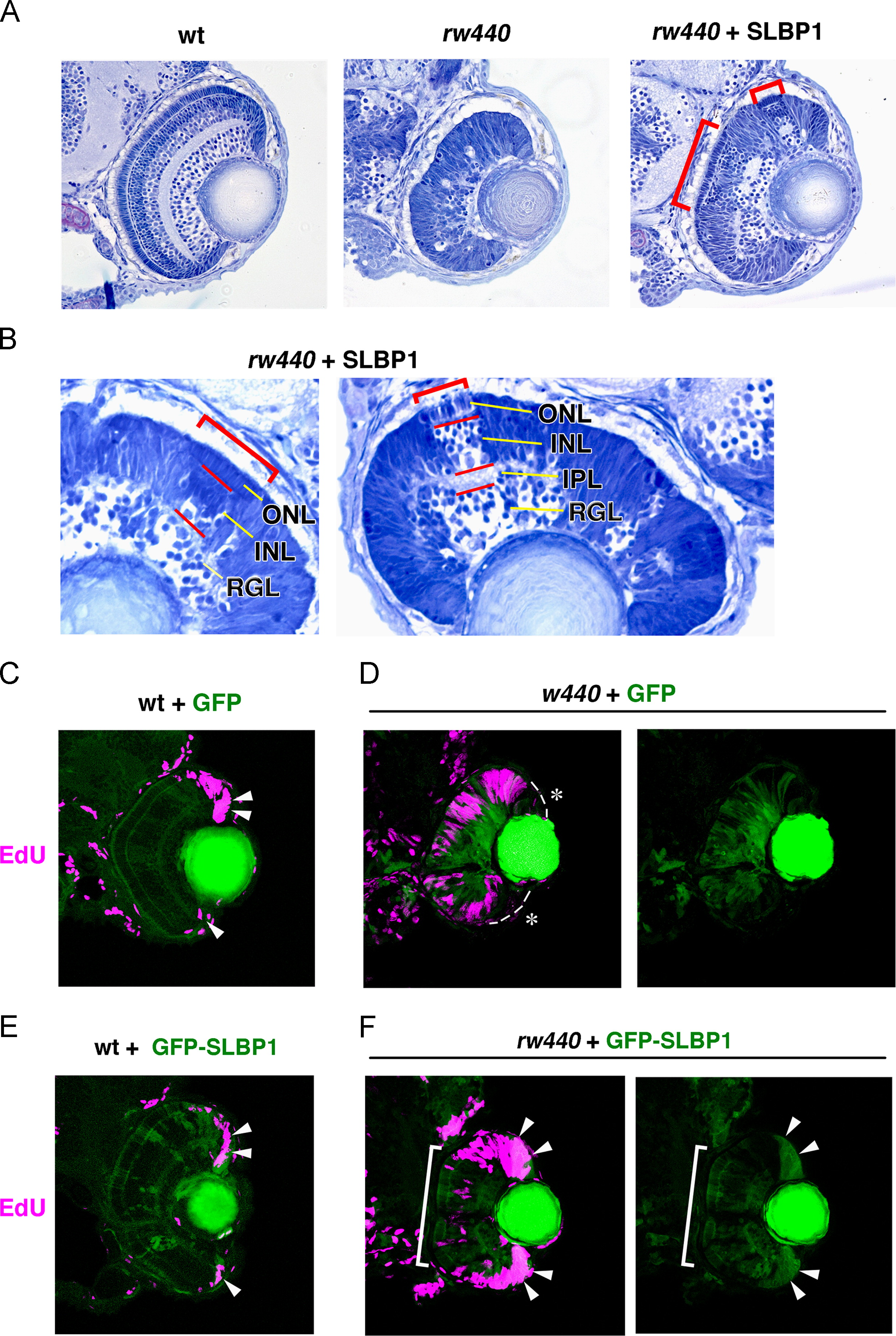
Fig. 6
Introduction of SLBP1 rescues cell proliferation and neurogenic defects in the rw440 mutant retina. (A) Sections of wild-type (left), rw440 mutant (middle), and rw440 mutant retinas injected with Tol2[EF1α: SLBP1] (right). Retinal lamination is recovered in columns of the rw440 mutant retinas injected with Tol2[EF1α: SLBP1] (red brackets). (B) High magnification of the rw440 mutant retinas injected with Tol2[EF1α: SLBP1]. Red brackets indicate restored retinal lamination. (C and D) EdU incorporation (magenta) of wild-type (C) and the rw440 mutant (D) retinas injected with Tol2[EF1α: GFP]. EdU incorporation is observed in the CMZ of wild-type retinas (arrowheads, C), but not in the CMZ of rw440 mutant retinas (asterisks, D). The right panel of (D) is the green channel. (E and F) EdU incorporation (magenta) of wild-type (E) and rw440 mutant (F) retinas injected with Tol2[EF1α: GFP-SLBP1]. Expression of GFP-SLBP1 did not have effects on EdU incorporation in wild-type retinal CMZ (arrowheads, E). In the rw440 mutant, expression of GFP-SLBP1 restores EdU incorporation in the retinal CMZ (arrowheads, F), and suppresses EdU incorporation in the central retina (bracket, F). The right panel of (F) is the green channel. RGL, RGC layer; IPL, inner plexiform layer; INL, inner nuclear layer; ONL, outer nuclear layer.
Reprinted from Developmental Biology, 394(1), Imai, F., Yoshizawa, A., Matsuzaki, A., Oguri, E., Araragi, M., Nishiwaki, Y., Masai, I., Stem-loop binding protein is required for retinal cell proliferation, neurogenesis, and intraretinal axon pathfinding in zebrafish, 94-109, Copyright (2014) with permission from Elsevier. Full text @ Dev. Biol.

