
|
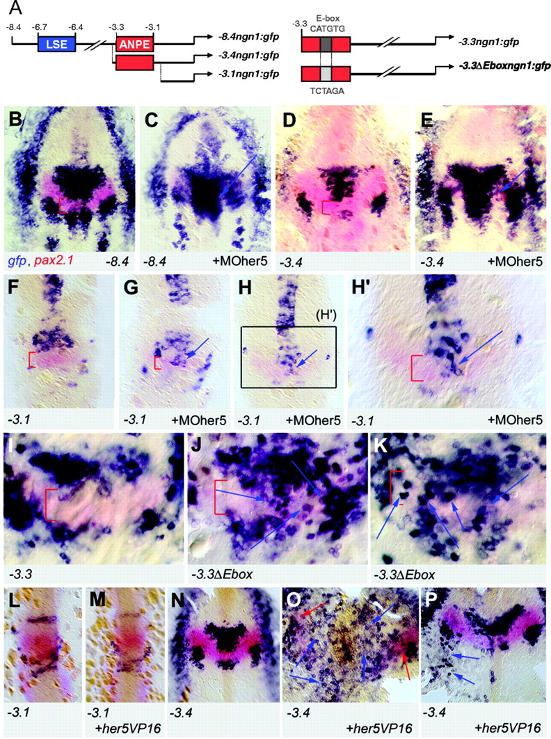
Fig. 6 An E-box contained within the ANPE element of the ngn1 gene is the major Her5 response element. (A) ngn1 transgenic reporter lines (left panel) (Blader et al., 2003) and reporter constructs used in transient assays (right panel) used to locate the response elements to Her5 within the ngn1 enhancer. (B-H2) Expression of gfp (revealed by in situ hybridisation, blue staining) and pax2.1 (red staining, used to located the IZ) in the following transgenic lines: –8.4ngn1:gfp (B,C),– 3.4ngn1:gfp (D,E), –3.1ngn1:gfp (F-H2) upon injection of MOher5 (C,E,G-H2) or in non-injected siblings (B,D,F). All panels are flat-mounted embryos, anterior towards the top, at the three-somite (B-E) and eight-somite (F-H2) stages; red brackets indicate the IZ. Two different embryos are shown for injection in the –3.1 line (G,H); H2 is a highly magnified view of the area boxed in H. Note that gfp expression is strongly induced across the IZ upon block of Her5 activity in the –8.4 and –3.4 lines, in a manner similar to endogenous ngn1 expression, but that the response of the –3.1 transgene is minor and restricted to a few cells at the ventral midline. (I-K) Expression of gfp (blue) and pax2.1 (red) in founder embryos injected with –3.3ngn1:gfp (I) and –3.3 Eboxngn1:gfp (J,K, two different embryos are shown). Both constructs carry SceI sites at their extremities and were co-injected with the meganuclease enzyme to trigger early integration (Thermes et al., 2002). Note the large number of ectopic gfp-positive cells in the entire medial IZ domain in embryos expressing the mutated construct without blocking Her5 activity, demonstrating that the E-box located within the ANPE is the major element mediating ngn1 repression at the IZ in vivo. (L-P) Expression of gfp (blue) and pax2.1 (red) in transgenic embryos (lines indicated bottom left). Uninjected embryos (L,N); embryos injected with her5VP16 capped RNA (M,O,P). Embryos are observed at the eight-somite (L,M) and one-somite (N-P) stage. Note that gfp expression in –3.1ngn1:gfp embryos is unperturbed by Her5VP16 (M), while ectopic expression is evident in –3.4ngn1:gfp embryos (two examples shown in O,P, blue arrows indicate ectopic gfp-positive cells, red arrows indicate pax2.1 expression in O).