
Fig. 7
- ID
- ZDB-IMAGE-141230-10
- Publication
- Amacher et al., 2002 - The zebrafish T-box genes no tail and spadetail are required for development of trunk and tail mesoderm and medial floor plate
- All Figures
- Figures for Amacher et al., 2002
|
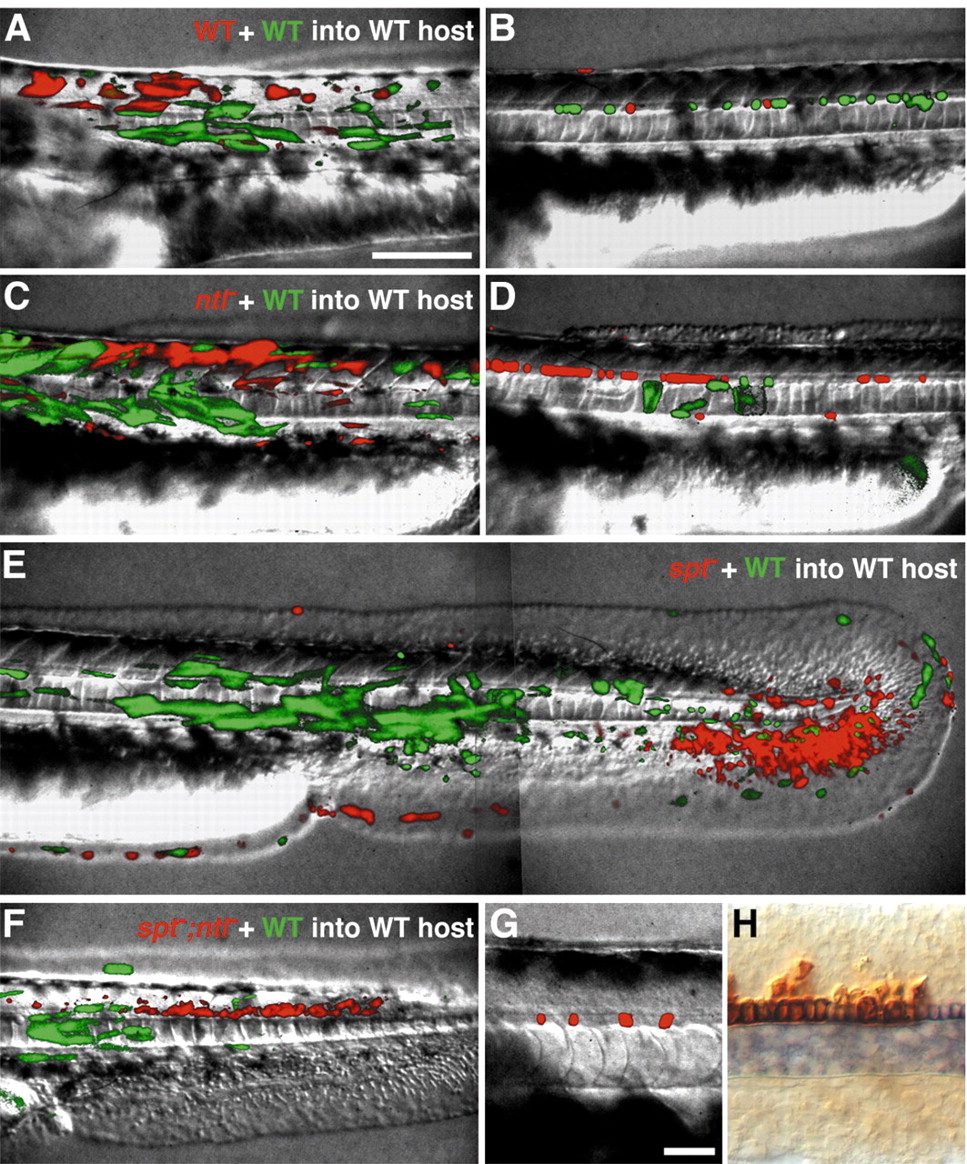
Fig. 7 spt and ntl together are required cell-autonomously in mesodermal cells, but neither gene is required in medial floor plate cells. Genetic mosaic embryos were generated by isochronic transplantation of blastula cells from a rhodamine-labeled donor embryo derived from an intercross of two heterozygous spt;ntl carriers, together with blastula cells from a fluorescein-labeled wild-type donor, into the presumptive mesoderm region of a wild-type host embryo (see Materials and Methods for details). Shown are live embryos at approximately 36 hpf with rhodamine- and fluorescein-labeled cells visualized in red and green, respectively (A-G), and an embryo fixed at 22 hpf and processed to detect shh expression (blue) and a co-injected biotinylated-dextran lineage tracer (brown) (H). In A-F, the fluorescein-labeled cells are wildtype, and rhodamine-labeled cells are either wildtype (A,B), ntl– (C,D), spt– (E), or spt–;ntl– (F,G). Cells doubly homozygous for spt and ntl mutations never make mesoderm, but instead contribute to non-neural ectoderm, spinal cord, and MFP (F,G). Some genetic mosaics containing spt–;ntl– cells were fixed at approximately 22 hpf and processed to detect shh transcripts (blue) and the co-injected biotinylated-dextran lineage tracer (brown) to show that donor-derived spt–;ntl– floor plate cells express shh, a MFP marker (H). Scale bars: 100 μm (A-F), 25 μm (G,H).